Tumor image focus area prediction analysis method and system and terminal equipment

A predictive analysis and tumor imaging technology, applied in the field of deep learning, can solve problems such as unsatisfactory classification prediction results, achieve the effects of improving classification prediction results, realizing feature enhancement, and removing redundant features of image data

Pending Publication Date: 2021-05-14
JIANGSU UNIV
View PDF0 Cites 4 Cited by
  • Summary
  • Abstract
  • Description
  • Claims
  • Application Information

AI Technical Summary

Problems solved by technology

However, no matter what kind of network structure is used, it is classified through the single data source of image, but due to the individual differences of patients, it is difficult to judge through the one-way data source, so the current classification prediction effect is not ideal

Method used


Image

  • Tumor image focus area prediction analysis method and system and terminal equipment
  • Tumor image focus area prediction analysis method and system and terminal equipment
  • Tumor image focus area prediction analysis method and system and terminal equipment

Examples


Embodiment Construction

[0045] The present invention will be further described below in conjunction with the accompanying drawings and specific embodiments, but the protection scope of the present invention is not limited thereto.

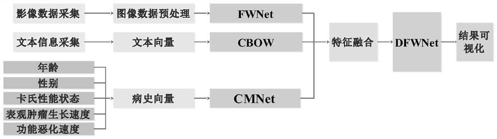
[0046] Such as figure 1 As shown, a method for predicting and analyzing lesion areas of tumor images, the specific steps are as follows:

[0047]Step (1), collect image data, diagnosis text and medical history data, and establish image, diagnosis text and medical history database

[0048] 1) The image data is obtained from the hospital, and the tumor image slices are obtained by precise and high-quality computed tomography (CT); the image data should include data from different cases, different types of lesions, and data from different hospitals; , each slice must be named with a standardized name, the name should be lesion type_period_serial number.dcm, for example, the slice with serial number 32 of metastatic liver adenocarcinoma should be named MET_PS_32.dcm; Afterw...


PUM


Abstract

The invention provides a tumor image focus area prediction analysis method and system and terminal equipment, and belongs to the field of deep learning. The method comprises the steps of collection of image data, diagnosis text and medical history data and prediction analysis of a tumor focus area, and specifically comprises the steps of preprocessing the collected data, and extracting image features through a constructed fusion weighted extraction network model; constructing a one-dimensional vector by using the medical history characteristics of a patient, namely age, gender, Karnofsky performance state, apparent tumor growth speed and function deterioration speed, and extracting the medical history characteristics by using a constructed text characteristic extraction network model of a dynamic convolution kernel; and after text features obtained by using the CBOW network model are fused with medical history features and image features, performing focus area prediction analysis through the constructed double-layer weighted prediction analysis network model. The method can significantly improve the classification prediction effect of the tumor image.

Description

technical field [0001] The invention belongs to the field of deep learning, and in particular relates to a method, system and terminal equipment for predicting and analyzing lesion areas of tumor images. Background technique [0002] In recent years, people's illnesses have shown an upward trend, and the main causes of modern people's illnesses are divided into three categories: one is caused by people's psychological factors, endocrine disorders caused by people's gradually increasing work pressure, abnormal mood swings, immune Second, it is caused by physical factors, such as irregular life schedule, frequent staying up late, unhealthy living habits such as smoking and drinking, ionizing radiation caused by nature or industrial production and medicine, etc. The occurrence of malignant tumors; the third is caused by organic chemical factors, such as benzopyrene in tobacco, which has obvious carcinogenic effects. Aflatoxin can cause lung cancer, skin diseases, and liver canc...

Claims


Application Information

Patent Timeline
Patent Type & Authority Applications(China)
IPC IPC(8): G06K9/62G06N3/04G06N3/08G16H50/20G16H50/30G16H50/70
CPCG06N3/084G16H50/20G16H50/30G16H50/70G06V2201/032G06N3/045G06F18/2415G06F18/253G06F18/214
Inventor 宋余庆毛静怡刘哲
Owner JIANGSU UNIV
Why Eureka
Try Eureka