A kind of medical radioactive silicon dioxide microsphere and its preparation method and application
A silicon dioxide and radioactive technology, applied in the direction of radioactive carriers, drug combinations, antineoplastic drugs, etc., can solve the problems of large side effects, poor accuracy, and large normal tissue damage, and achieve less radioactive waste, low production costs, and reduce radioactivity radiation effect
- Summary
- Abstract
- Description
- Claims
- Application Information
AI Technical Summary
Problems solved by technology
Method used
Image
Examples
Embodiment 1
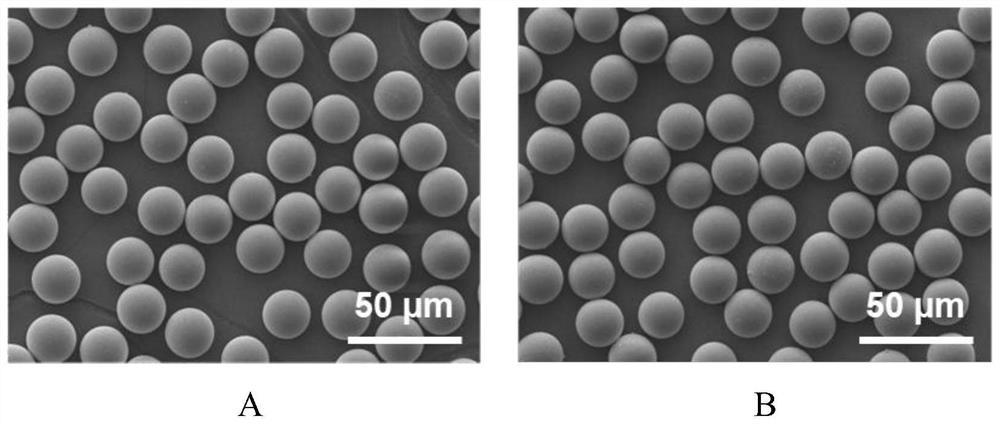
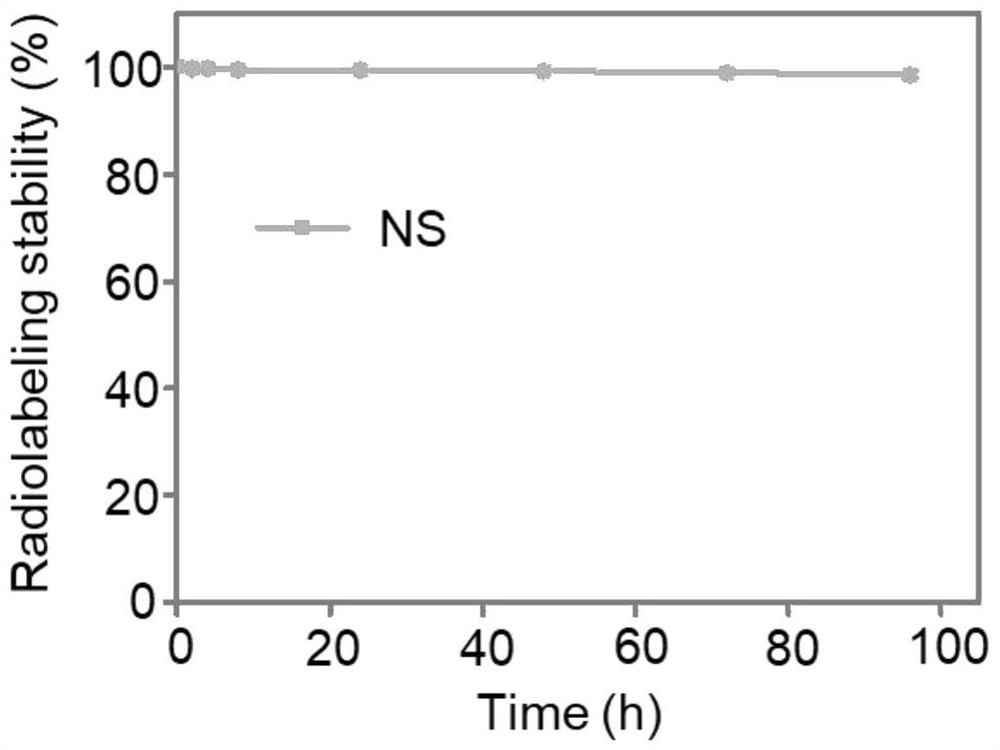
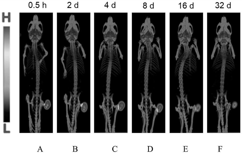
[0032] Experimental equipment: one ten-thousandth balance (ME204, Mettler-Toledo Shanghai Co., Ltd.); ultrapure water system (DirectQ5, MerckMillipore, USA); constant temperature mixer (YY10, Shanghai Yunyan Instrument Co., Ltd.); radioactivity Activity meter (FJ-391A4, Beijing Nuclear Instrument Factory); gamma radioimmunoassay counter (LB2111, German BERTHOLD company); scanning electron microscope (EVO18, German Carl Zeiss company); SPECT / CT imaging system (Milabs, Hong Kong Merrill Lynch Group) Co., Ltd.); fluorescence microscope (IX73, Japan Olympus Company); commonly used glass and surgical utensils.
[0033] Experimental reagent: silica microspheres (Suzhou Zhiyi Microsphere Technology Co., Ltd.); 177 LuCl 3 Solution (Sichuan Xinke Pharmaceutical Co., Ltd.); potassium hydroxide (Shanghai Aladdin Biochemical Technology Co., Ltd.); phosphoric acid (Shanghai Aladdin Biochemical Technology Co., Ltd.); normal saline (Shanghai Yuanye Biotechnology Co., Ltd.); Ma Lin (Shangha...
Embodiment 3
[0052] Disperse 8mg of silica microspheres in 10mL of pure water, add 1Ci medical radionuclide yttrium chloride [ 90 YCl 3 ] solution, at room temperature, shake in a constant temperature mixer for 10 minutes to prepare a silica microsphere mixture. Under shaking at room temperature, add 0.025 mL of sodium carbonate-sodium hydroxide buffer solution (pH = 12) dropwise to the above silica microsphere mixture, and continue shaking for 30 minutes. After solid-liquid separation, wash with pure water for 4 times, after draining, yttrium carbonate [ 90 YCO 3 ] Medical radioactive silica microspheres.
Embodiment 4
[0054] Disperse 10 mg of silica microspheres in 0.6 mL of pure water, add 0.2 Ci of medical radionuclide yttrium chloride [ 90 YCl 3 ] solution, 0.1Ci medical radionuclide lutetium chloride [ 177 LuCl 3 ] solution, 0.4Ci medical radionuclide strontium chloride [ 89 SrCl 2 ], at room temperature, shake in a constant temperature mixer for 10 minutes to prepare a mixture of silica microspheres. Under shaking at room temperature, add 0.2 mL of potassium chloride-sodium hydroxide (pH = 10) dropwise to the above silica microsphere mixture, continue shaking for 30 minutes, and wash with pure water for 5 times after solid-liquid separation , after draining, the resulting 90 Y, 177 Lu, 89 Hydroxide silica microspheres of various medical radionuclides of Sr.
PUM
 Abstract
Description
Claims
Application Information
 - R&D
- Intellectual Property
- Life Sciences
- Materials
- Tech Scout
- Unparalleled Data Quality
- Higher Quality Content
- 60% Fewer Hallucinations
Browse by: Latest US Patents, China's latest patents, Technical Efficacy Thesaurus, Application Domain, Technology Topic, Popular Technical Reports.
© 2025 PatSnap. All rights reserved.Legal|Privacy policy|Modern Slavery Act Transparency Statement|Sitemap|About US| Contact US: help@patsnap.com


