Myocardial fine-grained division method and system

A fine-grained, myocardial technology, applied in image analysis, image enhancement, instruments, etc., can solve the problem of not giving the method for determining the junction point of the left and right ventricles, and achieve the effect of facilitating computer-aided diagnosis.

Pending Publication Date: 2021-05-28
SHANGHAI UNIV
View PDF0 Cites 0 Cited by
  • Summary
  • Abstract
  • Description
  • Claims
  • Application Information

AI Technical Summary

Problems solved by technology

[0003] The American Heart Association's myocardial 16-segment method can be generally used in cardiac MRI short-axis images, but because there is no method for determining the junction point of the left and right ventricles, it is more suitable for human working maps

Method used


Image

  • Myocardial fine-grained division method and system
  • Myocardial fine-grained division method and system
  • Myocardial fine-grained division method and system

Examples


Embodiment Construction

[0050] The following will clearly and completely describe the technical solutions in the embodiments of the present invention with reference to the accompanying drawings in the embodiments of the present invention. Obviously, the described embodiments are only some of the embodiments of the present invention, not all of them. Based on the embodiments of the present invention, all other embodiments obtained by persons of ordinary skill in the art without making creative efforts belong to the protection scope of the present invention.

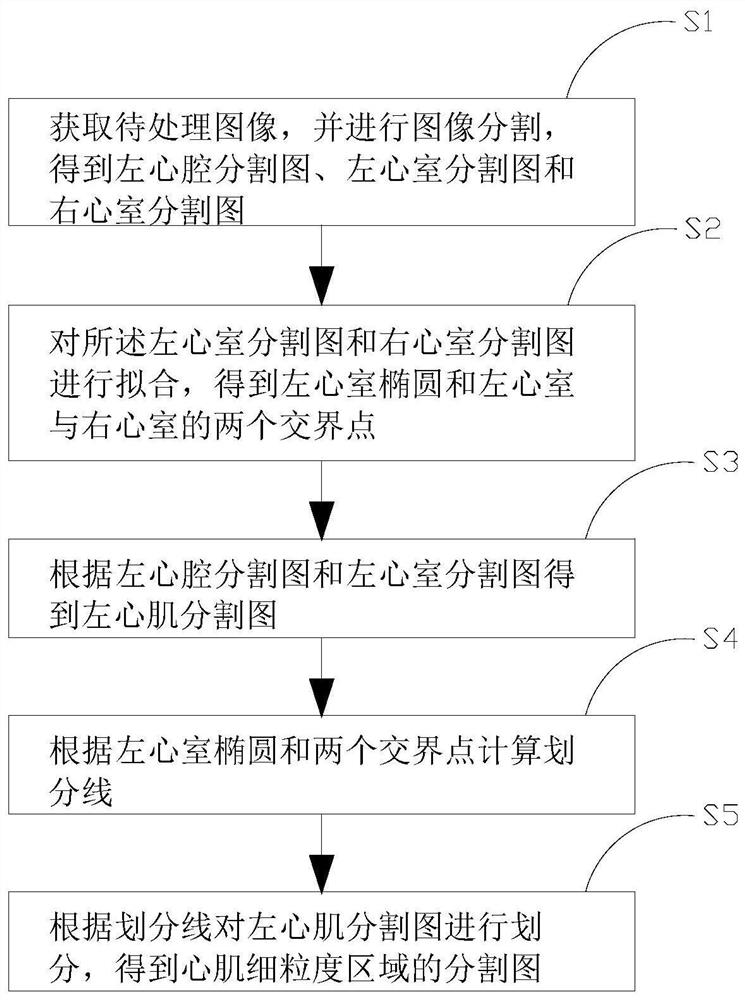
[0051] Such as figure 1 As shown, the embodiment of the present invention proposes a myocardial fine-grained classification method, including the following steps:

[0052] A method for fine-grained division of myocardium, comprising the following steps:

[0053] S1. Acquire the image to be processed, and perform image segmentation to obtain a segmentation map of the left ventricle, a segmentation map of the left ventricle, and a segmentation map...


PUM


Abstract

The invention provides a myocardial fine-grained division method and system, and the method comprises the steps: obtaining a to-be-processed image, and carrying out the image segmentation, and obtaining a left heart cavity segmentation image, a left ventricle segmentation image, and a right ventricle segmentation image; s2, fitting the left ventricle segmentation image and the right ventricle segmentation image to obtain a left ventricle ellipse and two junction points of the left ventricle and the right ventricle; s3, obtaining a left cardiac muscle segmentation map according to the left cardiac cavity segmentation map and the left ventricle segmentation map; s4, calculating a division line according to the left ventricular ellipse and the two junction points; and S5, dividing the left myocardial segmentation map according to the division line to obtain a segmentation map of the myocardial fine-grained region. According to the myocardial fine-grained division method and system, a machine can calculate the division line according to the left ventricular ellipse and the two junction points to perform myocardial fine-grained division, so that automatic computer-aided diagnosis is facilitated.

Description

technical field [0001] The present invention relates to the technical field of myocardial tissue examination, in particular to a method and system for dividing myocardial fine grains. Background technique [0002] T1 mapping technology is a good non-invasive myocardial tissue examination method, which can play a very good role in the diagnosis, examination and prognosis of various cardiovascular diseases. In order to accurately describe the abnormal region of the cardiac T1mapping image, it is necessary to divide the myocardium into multiple regions. The 16-segment method of the American Heart Association. It divides the heart into three equal parts along the long axis of the heart: the apex, the heart, and the base of the heart (apical, mid, and basal). The base of the heart is equally divided into 6 regions, a total of 16 regions. [0003] The American Heart Association's myocardial 16-segment method can be generally used in cardiac MRI short-axis images, but because it...

Claims


Application Information

Patent Timeline
IPC IPC(8): G06T7/00G06T7/11
CPCG06T7/0012G06T2207/10088G06T2207/30048G06T7/11
Inventor 沈文枫王陈星宇
Owner SHANGHAI UNIV
Why Eureka
Try Eureka