Time-resolved fluorescence immunoassay method of content of sH2a in serum, and detection kit thereof

A time-resolved, immunofluorescence technology, applied in the field of protein detection, can solve the problems of narrow linear range, long detection time, high detection background, and achieve the effect of wide linear range, short detection time and high accuracy.

Inactive Publication Date: 2015-07-29
JIANGSU MICRODIAG BIOMEDICINE TECH CO LTD
View PDF3 Cites 6 Cited by
  • Summary
  • Abstract
  • Description
  • Claims
  • Application Information

AI Technical Summary

Problems solved by technology

[0003] In the previous research work, the inventor invented a sH2a quantitative detection kit (CN103336125A), which uses double-antibody sandwich enzyme-linked immunosorbent assay to quantitatively detect sH2a, but the invention still has a narrow linear range and a high detection background , the detection takes a long time and other problems, and needs to be further improved

Method used


Image

  • Time-resolved fluorescence immunoassay method of content of sH2a in serum, and detection kit thereof
  • Time-resolved fluorescence immunoassay method of content of sH2a in serum, and detection kit thereof
  • Time-resolved fluorescence immunoassay method of content of sH2a in serum, and detection kit thereof

Examples


Embodiment 1

[0044] Embodiment 1, expression, purification and identification of sH2a recombinant protein

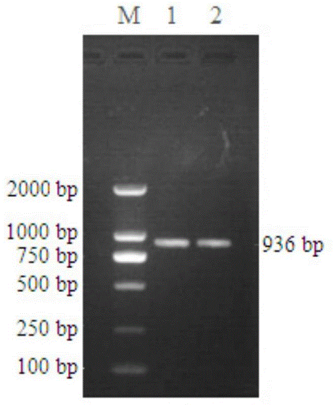
[0045] Using liver cancer tissue cDNA as template, 5'-CGGGATCCATGGCCAAGGACTTTCAAGATATCCA-3' (SEQ ID No.1) and 5'-CGGAATTCTCAGGCCACCTCGCCGGT-3' (SEQ ID No.2) as primers, PCR amplified sH2a gene fragment ( figure 1 ), after gel cutting, recovery and purification, it was connected to the cloning vector pCR2.1 and sequenced for identification, and then the sH2a gene fragment with the correct sequence identification was cloned into the expression vector pET28a(+), the restriction sites were BamH I and EcoR I, The recombinant plasmid pET28a-sH2a was obtained, which contained 6×HIS tag, which was convenient for subsequent protein purification.

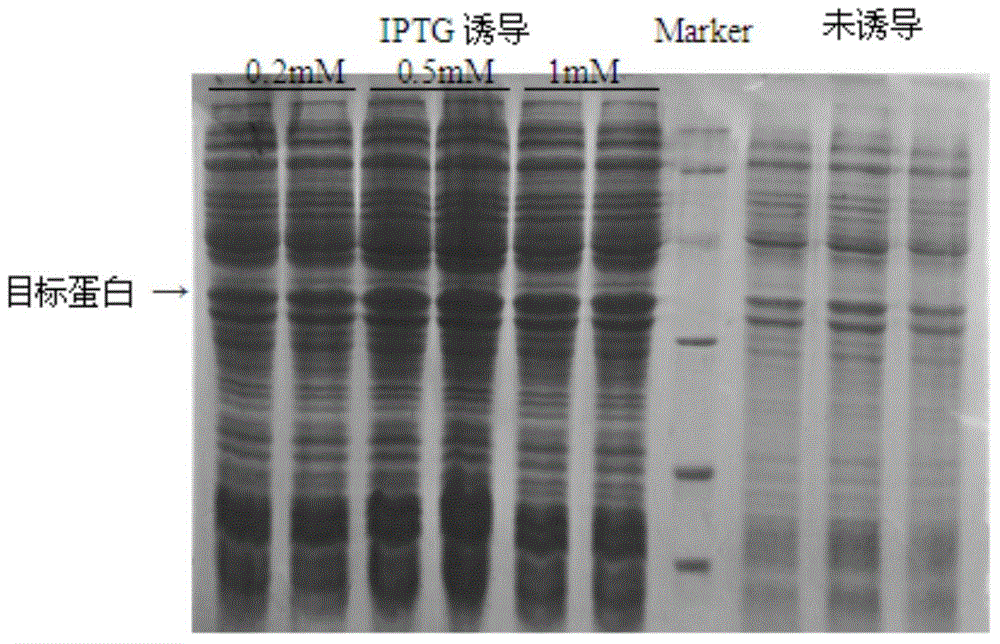
[0046] The recombinant plasmid pET28a-sH2a was transformed into Escherichia coli BL21(DE3), and the successfully transformed clones were picked and cultured at 37°C until the OD600nm value was about 0.6, and 0.2, 0.5, and 1 mM IPTG were added respec...

Embodiment 2

[0048] Embodiment 2, preparation of anti-sH2a monoclonal antibody

[0049] Two 6-week-old BALB / c female mice were taken and immunized according to the following steps:

[0050] First immunization: Mix the sH2a recombinant protein solution with Freund’s complete adjuvant at a volume ratio of 1:1, fully emulsify, and inject 0.1ml subcutaneously at two points on the back of the mouse (30 μg of sH2a recombinant protein per mouse);

[0051] The second immunization: after an interval of 14 days, mix the sH2a recombinant protein solution with Freund’s incomplete adjuvant at a volume ratio of 1:1, fully emulsify, and inject 0.1ml (sH2a recombinant Protein 50μg / piece);

[0052] The third immunization: after an interval of 14 days, mix the sH2a recombinant protein solution with Freund’s incomplete adjuvant at a volume ratio of 1:1, fully emulsify, and inject 0.1ml subcutaneously at the back of the shoulder and abdomen on both sides of the mouse (sH2a recombinant protein 50μg / piece); ...

Embodiment 3

[0057] Example 3, paired screening of anti-sH2a monoclonal antibodies

[0058] The above 9 strains of purified anti-sH2a monoclonal antibodies were respectively coated in solid-phase microwell plates, and the other 8 strains of anti-sH2a monoclonal antibodies labeled with Eu3+ except themselves were used for pairwise pairing experiments. The results showed that anti-sH2a monoclonal antibodies 2D4H5 and 4A7D8 could be paired to establish a reaction system (Table 2).

[0059] Table 2s Screening of H2a paired monoclonal antibodies (double-antibody sandwich method, coated with 2D4H5)

[0060]


PUM


Abstract

The invention discloses a time-resolved fluorescence immunoassay method of the content of sH2a in serum. The method comprises the following steps: adding an sH2a substandard substance or a serum sample to be detected into an anti-sH2a monoclonal antibody coated solid phase material, incubating, washing, adding a lanthanide element ion labeled anti-sH2a monoclonal antibody capable of being paired to the anti-sH2a monoclonal antibody, incubating, washing, adding a dissociation enhancement liquid, shaking, determining the fluorescence intensity, drafting a standard curve according to the determination result of the sH2a substandard substance, and calculating the content of sH2a in the serum sample to be detected according to the standard curve. The invention also discloses a time-resolved fluorescence immunoassay kit of the content of sH2a in the serum. The detection method and the kit have the advantages of wide linear range, zero background, good stability, high sensitivity, strong specificity, high accuracy and short detection time, can be used for detecting the content of sH2a in hepatopathy patient serum, and is convenient for clinic assisted diagnosis of hepatopathy.

Description

technical field [0001] The invention belongs to the technical field of protein detection, and relates to a protein quantitative detection method and a detection kit. Background technique [0002] sH2a is a soluble secreted protein expressed specifically in the liver, with a molecular weight of about 35KD. It is homologous to the H2 subunit of another asialic acid receptor protein (ASGPR), which is also expressed only in hepatocytes, but studies have shown that sH2a is not involved in the assembly of the ASGRP H2 subunit. After the precursor protein of sH2a is expressed in hepatocytes, the endoplasmic reticulum is cut and removed about 5 amino acids to become sH2a, which is released outside the cell and enters the blood circulation. The sH2a level in the peripheral blood of normal people maintains a high level, but the sH2a content decreases significantly in patients with liver diseases such as liver injury, hepatitis, and liver fibrosis. Auxiliary diagnosis of liver injury...

Claims


Application Information

Patent Timeline
Patent Type & Authority Applications(China)
IPC IPC(8): G01N33/68
CPCG01N33/577G01N33/533
Inventor 王弢秦勇周小进陈菲
Owner JIANGSU MICRODIAG BIOMEDICINE TECH CO LTD
Why Eureka
Try Eureka