
New coronal pneumonia focus segmentation method based on feature fusion deep supervision U-Net

A feature fusion and lesion technology, applied in the field of new coronary pneumonia lesion segmentation based on feature fusion deep supervision U-Net, can solve the problem of different importance, achieve the effect of improving accuracy and improving work efficiency

Pending Publication Date: 2021-06-11
NORTHEASTERN UNIV
View PDF2 Cites 6 Cited by
  • Summary
  • Abstract
  • Description
  • Claims
  • Application Information

AI Technical Summary

Problems solved by technology

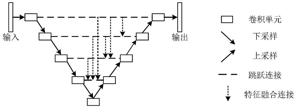
However, the structure only uses single-level single-scale features, and the importance of different levels of features is different for different data.

Method used


Image

  • New coronal pneumonia focus segmentation method based on feature fusion deep supervision U-Net
  • New coronal pneumonia focus segmentation method based on feature fusion deep supervision U-Net
  • New coronal pneumonia focus segmentation method based on feature fusion deep supervision U-Net

Examples


Embodiment Construction

[0037] The present invention will be further described below in conjunction with the accompanying drawings and specific embodiments.

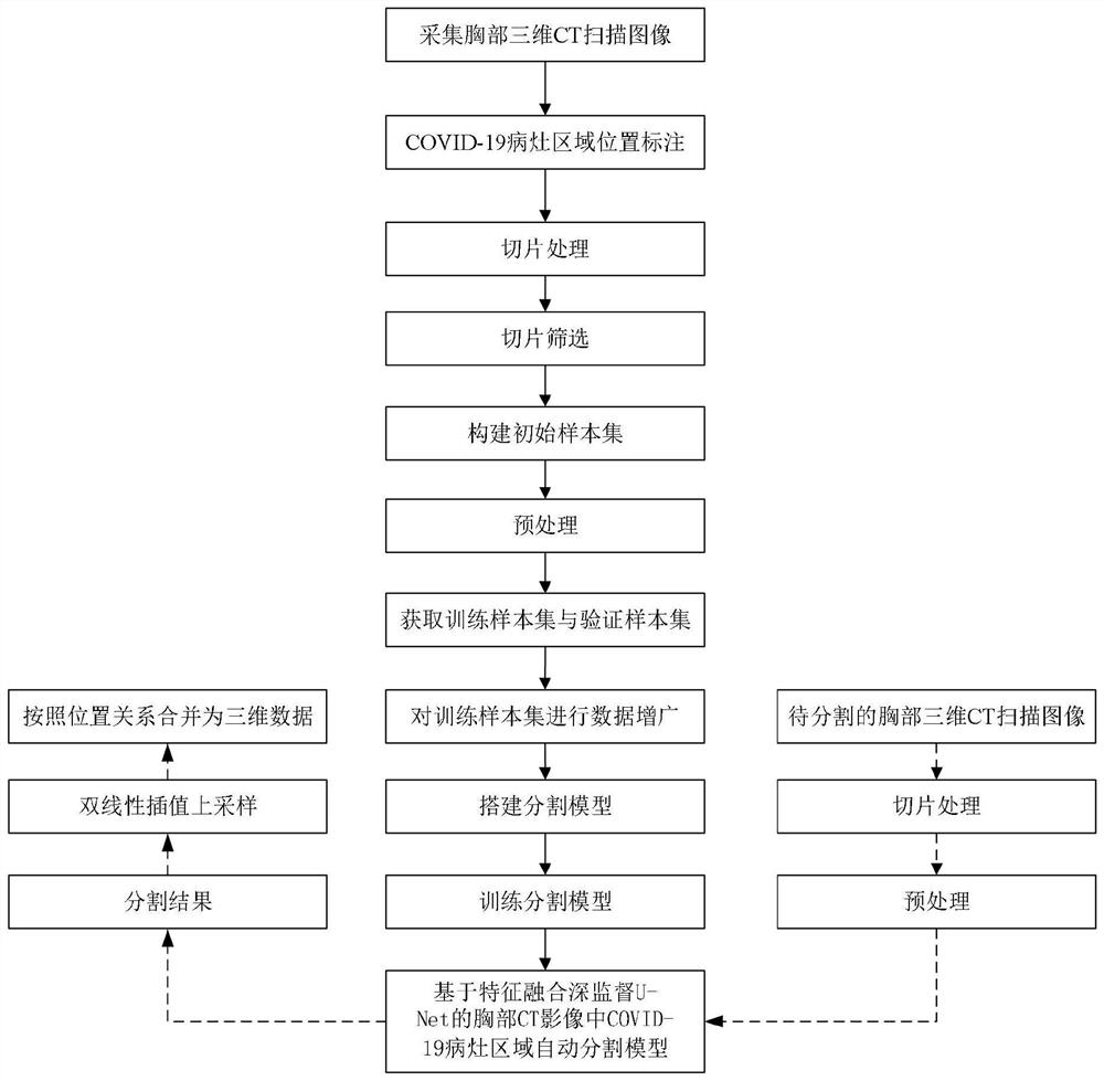
[0038] Such as figure 1 As shown, the new coronary pneumonia lesion segmentation method based on feature fusion deep supervision U-Net of the present invention includes the following steps:

[0039] Step 1: Obtain an initial sample set

[0040] Collect the chest 3D CT scan images of multiple patients diagnosed with COVID-19, mark the location of the COVID-19 lesion area in the chest 3D CT scan images, perform slice processing on the chest 3D CT scan images, and screen out those that do not contain COVID-19. For the slices of 19 lesions, all the slices left after the screening of each patient and the labeled data corresponding to each slice are used as an initial sample to obtain an initial sample set.

[0041] Step 2: Preprocessing the initial sample set

[0042] In this embodiment, step 2 includes the following steps:

[0043] Step 2.1: Pe...


PUM


Abstract

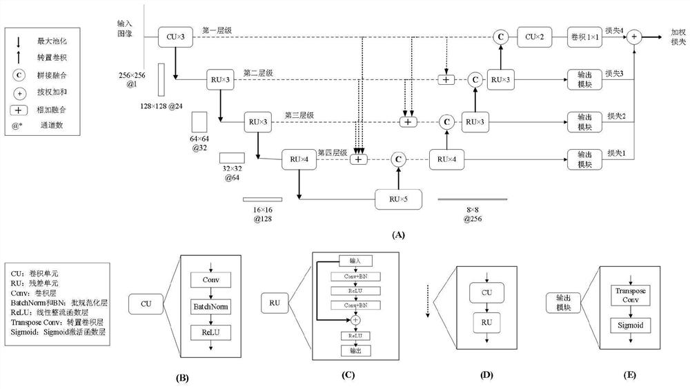
The invention relates to the technical field of medical image segmentation, and provides a new coronal pneumonia focus segmentation method based on feature fusion deep supervision U-Net, and the method comprises: 1, obtaining an initial sample set; 2, preprocessing the initial sample set; 3, obtaining a training sample set and a verification sample set; 4, performing data augmentation on the training sample set; 5, building a COVID-19 lesion area automatic segmentation model in the chest CT image based on feature fusion deep supervision U-Net, wherein the automatic segmentation model comprises an encoder, a decoder, jump connections, feature fusion blocks and deep supervision branches, adding the feature fusion blocks between the jump connections of different levels, and adding the deep supervision branches to the decoder part; 6, training a segmentation model; and 7, segmenting the chest three-dimensional CT scanning image to be segmented. According to the method, automatic segmentation of the COVID-19 lesion area in the chest CT can be realized, and the accuracy and rapidity of segmentation are improved.

Description

technical field [0001] The present invention relates to the technical field of medical image segmentation, in particular to a novel coronavirus pneumonia lesion segmentation method based on feature fusion deep supervision U-Net. Background technique [0002] At the end of 2019, the novel coronavirus (COVID-19) outbreak began worldwide. As of February 26, 2021, the number of confirmed cases worldwide has exceeded 100 million. On the one hand, each suspected case needs to be confirmed by RT-PCR testing. Although RT-PCR is the gold standard for diagnosis, the process is very time-consuming and the false-negative diagnosis is high. On the other hand, chest CT scans of COVID-19 patients usually show patchy ground-glass opacities on both sides of the lungs, which has been used as an important supplementary indicator for COVID-19 screening due to the high sensitivity of this feature. [0003] Chest CT examination also plays an important role in the follow-up evaluation of hospital...

Claims


Application Information

Patent Timeline
Patent Type & Authority Applications(China)
IPC IPC(8): G06T7/10G06K9/62G06N3/04G06N3/08
CPCG06T7/10G06N3/084G06T2207/10081G06T2207/30061G06N3/045G06F18/25G06F18/214
Inventor 曹鹏武博
Owner NORTHEASTERN UNIV
Learn More