Multi-modal liver tumor segmentation method based on MR and CT

A liver tumor, multi-modal technology, applied in the field of medical image processing, can solve the problems of low result accuracy and tumor segmentation accuracy, and achieve the effect of eliminating the mutual coupling relationship and improving the accuracy.

Pending Publication Date: 2021-08-06
北京精诊医疗科技有限公司
View PDF0 Cites 1 Cited by
  • Summary
  • Abstract
  • Description
  • Claims
  • Application Information

AI Technical Summary

Benefits of technology

The technical effect of this invention relates to improved image processing techniques for analyzing medical imagery data such as ultrasound scans or X-rays (XR) that help identify cancerous areas within an organism's body. This technology can be used alone or combined with other methods like histology analysis to improve diagnoses by identifying specific types of tissue abnormalities associated with certain diseases.

Problems solved by technology

This patents discuss how advances have happened during the past few decades when applying various techniques for analyzing medical images like x- ray fluorescence scans (XRF), sonar waves, magnetism resonance soundings, and other types of signals used in medicine. These improvements led to improved precision and safety in surgeries performed today. Additionally, these technical problem addressed through the patented technique involves developing a model system called Deep Learning Based Computer Programming System(DLPS). DLSPS can automatically extract relevant parts from existing datasets without requiring human input, making them easier than previously possible.

Method used


Image

  • Multi-modal liver tumor segmentation method based on MR and CT
  • Multi-modal liver tumor segmentation method based on MR and CT
  • Multi-modal liver tumor segmentation method based on MR and CT

Examples


Embodiment Construction

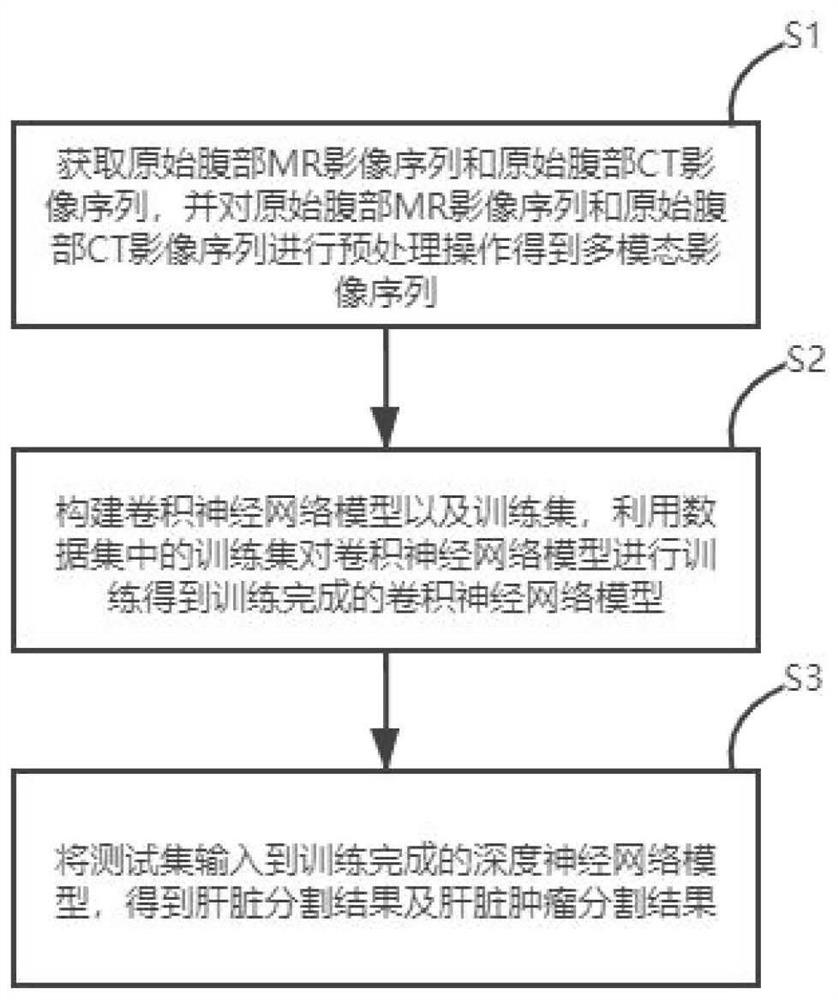
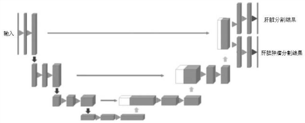
[0023] Below in conjunction with accompanying drawing and embodiment, technical solution of the present invention is described further:

[0024] This embodiment provides a multimodal liver tumor segmentation method based on MR and CT, the method comprising:

[0025] Step 1. Obtain the original abdominal MR image sequence and the original abdominal CT image sequence, and perform a preprocessing operation on the original abdominal MR image sequence and the original abdominal CT image sequence to obtain a multimodal image sequence.

[0026] In the embodiment of the present application, the preprocessing operation includes: performing bias field correction on the original abdominal MR image sequence to obtain the first MR image sequence. Uniformity is an unavoidable magnetic field effect, which will lead to uneven image range in the same tissue. In order to improve the accuracy of subsequent results, it is necessary to perform bias field correction on the original abdominal MRI im...


PUM


Abstract

The invention discloses a multi-modal liver tumor segmentation method based on MR and CT, and the method comprises the steps: firstly obtaining an original abdomen MR image sequence and an original abdomen CT image sequence, carrying out the preprocessing operation of the original abdomen MR image sequence and the original abdomen CT image sequence, obtaining a multi-modal image sequence, constructing a convolutional neural network model and a data set, and training the convolutional neural network model by using the training set in the data set to obtain a trained convolutional neural network model, and finally inputting the test set into the trained convolutional neural network model to obtain a segmentation result including the liver and the liver tumor. According to the method, the multi-modal image is used for liver and liver tumor segmentation, multi-modal information is fused, the precision of a final segmentation result is improved, liver and liver tumor segmentation tasks are carried out at the same time, and the mutual coupling relation between a liver segmentation result and a tumor segmentation result is eliminated.

Description


Claims


Application Information

Patent Timeline
Owner 北京精诊医疗科技有限公司
Why Eureka
Try Eureka