Ultrasonic contrast imaging method and device

A technology of contrast-enhanced ultrasound and imaging methods, which is applied in ultrasound/sound wave/infrasonic wave diagnosis, sound wave diagnosis, infrasonic wave diagnosis, etc. It can solve the problems of short time interval, unrealizable, incompletely consistent hepatic arteriography images, etc.

Pending Publication Date: 2021-08-31
SHENZHEN MINDRAY BIO MEDICAL ELECTRONICS CO LTD +1
View PDF0 Cites 1 Cited by
  • Summary
  • Abstract
  • Description
  • Claims
  • Application Information

AI Technical Summary

Problems solved by technology

In the current liver contrast-enhanced ultrasonography examination, although there is a certain time interval between the contrast images of the arterial phase and the portal venous phase, the time interval is very short. Microbubbles in the portal vein begin to appear, so the image quality of hepatic arteriography, especially in the late arterial stage, will be severely disturbed by microbubbles of contrast agent in the portal vein
According to the existing liver contrast-enhanced ultrasound imaging technology (intravenous injection of contrast agent), the hepatic arteriography image obtained by the doctor and the hepatic arteriography displayed by the method of injecting contrast agent directly through the hepatic artery in the operating room (invasive method) The image is not exactly the same
The current hepatic contrast-enhanced ultrasonography technology is still unable to realize pure hepatic arteriography imaging in the true sense, and cannot present the complete arterial enhancement process

Method used


Image

  • Ultrasonic contrast imaging method and device
  • Ultrasonic contrast imaging method and device
  • Ultrasonic contrast imaging method and device

Examples


Embodiment approach

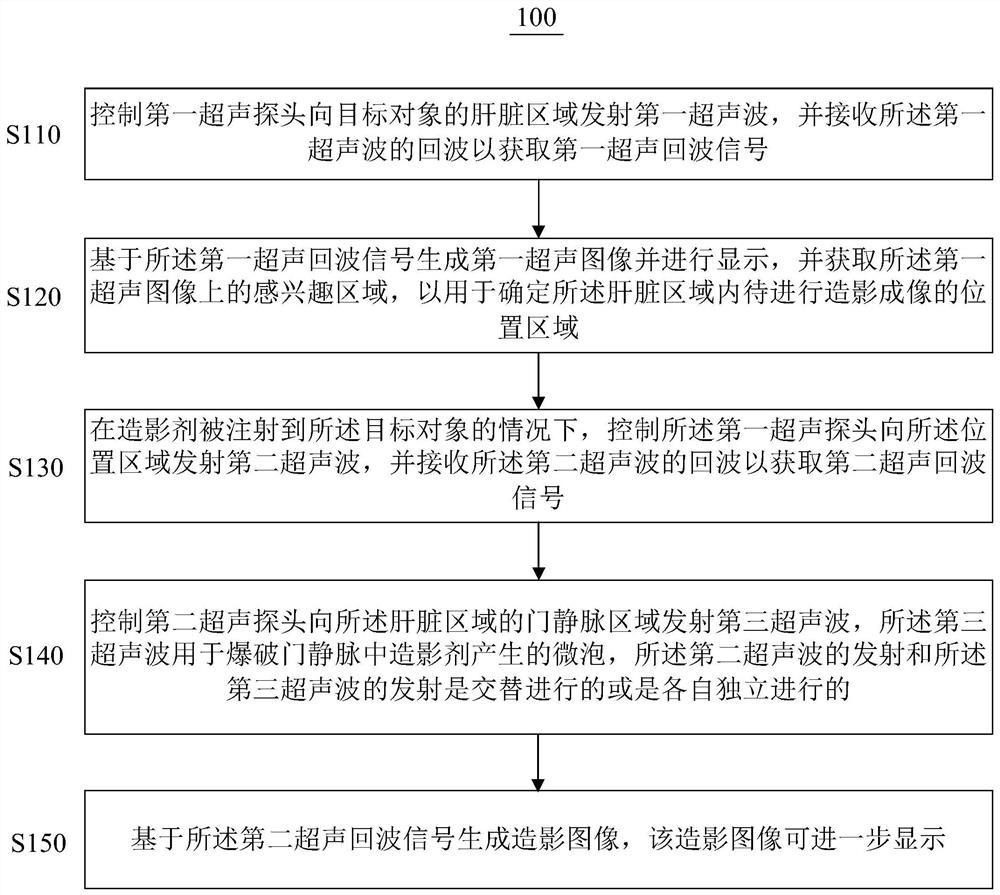
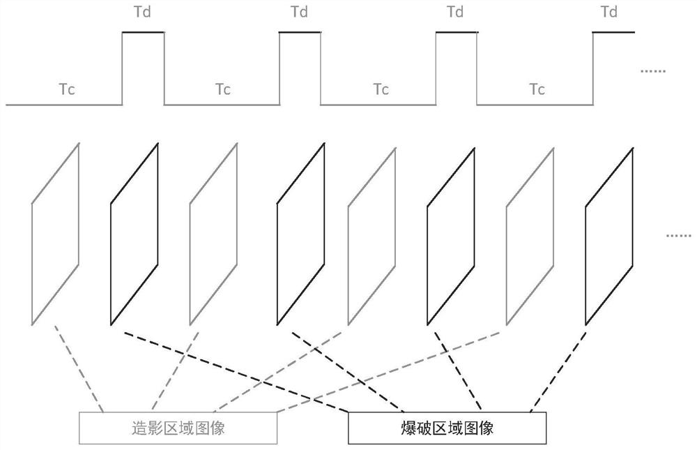
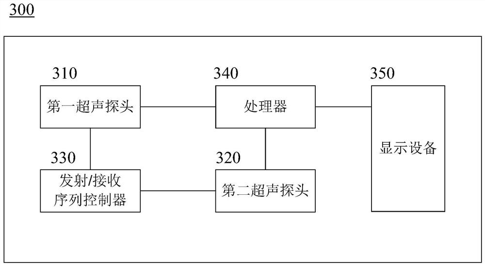
[0057] In addition, as mentioned above, the angiographic images of the arterial phase and the portal venous phase have a certain time interval in the appearance time sequence. Therefore, in one embodiment, the transmit / receive sequence controller 330 generally follows the injection of the contrast agent and satisfies the Under the preset conditions, the second ultrasound probe 320 is controlled to transmit a third ultrasound wave to the portal vein region of the liver region of the target object to burst the microbubbles generated by the contrast agent in the portal vein. In one example, the meeting the preset condition may include: timing from the injection of the contrast agent to a preset time. In this example, the preset time may be pre-defined according to the time interval between the angiographic images of the arterial phase and the portal venous phase in terms of appearance time, so that after a certain time of hepatic arteriography imaging, the microbubbles in the port...


PUM


Abstract

The invention provides an ultrasonic contrast imaging method and device. The method comprises the steps that a first ultrasonic probe is controlled to emit first ultrasonic waves to a liver area of a target object; a first ultrasonic image is generated based on a first ultrasonic echo signal, and an area of interest on the first ultrasonic image is acquired to determine a position area to be subjected to contrast imaging in the liver area; under the condition that a contrast agent is injected into the target object, the first ultrasonic probe is controlled to emit second ultrasonic waves to the position area; after the contrast agent is injected and when preset conditions are met, a second ultrasonic probe is controlled to emit third ultrasonic waves to a portal vein area of the liver area, wherein the third ultrasonic waves are used for bursting microbubbles generated by the contrast agent in the portal vein, and emission of the second ultrasonic waves and emission of the third ultrasonic waves are conducted alternately or conducted independently; and a contrast image is generated based on a second ultrasonic echo signal. According to the method and device, pure hepatic artery ultrasonic contrast imaging can be achieved.

Description

technical field [0001] The present invention relates to the technical field of ultrasound imaging, and more particularly to a method and device for contrast-enhanced ultrasound imaging. Background technique [0002] In modern medical imaging examinations, ultrasound technology has become the most widely used, most frequently used examination method and the fastest popularization and application of new technologies due to its high reliability, fast and convenient, real-time imaging, and repeatable examinations. The development of some new ultrasound technologies has further promoted the application of ultrasound imaging in clinical diagnosis and treatment. [0003] Ultrasonic waves will scatter when they encounter scatterers, and the intensity of the scattering is related to the size and shape of the scatterers and the difference in acoustic impedance with surrounding tissues. Although the blood contains tangible substances such as red blood cells, white blood cells, and pla...

Claims


Application Information

Patent Timeline
Patent Type & Authority Applications(China)
IPC IPC(8): A61B8/00
CPCA61B8/481
Inventor 费翔桑茂栋章希睿朱磊唐杰董多
Owner SHENZHEN MINDRAY BIO MEDICAL ELECTRONICS CO LTD
Why Eureka
Try Eureka