Liver tumor recognition method based on self-supervised dense convolutional neural network

A convolutional neural network and recognition method technology, applied in the field of liver tumor recognition based on self-supervised dense convolutional neural network, can solve the problems of insignificant early symptoms of HCC and insufficient training sample data, so as to reduce the amount of calculation and improve the calculation. Efficiency and improved accuracy

Pending Publication Date: 2021-09-07
SECOND AFFILIATED HOSPITAL OF XIAN MEDICAL UNIV
View PDF0 Cites 8 Cited by
  • Summary
  • Abstract
  • Description
  • Claims
  • Application Information

AI Technical Summary

Problems solved by technology

[0002] Liver cancer (hepatocellular carcinoma, HCC) is one of the malignant tumors with the highest mortality and morbidity in the world. Although surgical resection is the first choice for the treatment of liver cancer, the early symptoms of HCC are usually not obvious in clinical practice. In the middle and advanced stages, or due to conditions such as severe liver cirrhosis and abnormal liver function, less than 25% of the clinically discovered HCC can be surgically resected
U-net based on convolutional neural network technology is currently a well-known neural network framework in the medical field. Generate segmented images while ensuring the preservation of high-resolution information. Deep learning has the problem of insufficient training sample data in the field of medical images, because it is difficult to manually mark a large amount of data and require a relatively high accuracy rate.

Method used


Image

  • Liver tumor recognition method based on self-supervised dense convolutional neural network
  • Liver tumor recognition method based on self-supervised dense convolutional neural network
  • Liver tumor recognition method based on self-supervised dense convolutional neural network

Examples


Embodiment Construction

[0038] The present invention will be further described in detail below in conjunction with the accompanying drawings, which are explanations rather than limitations of the present invention.

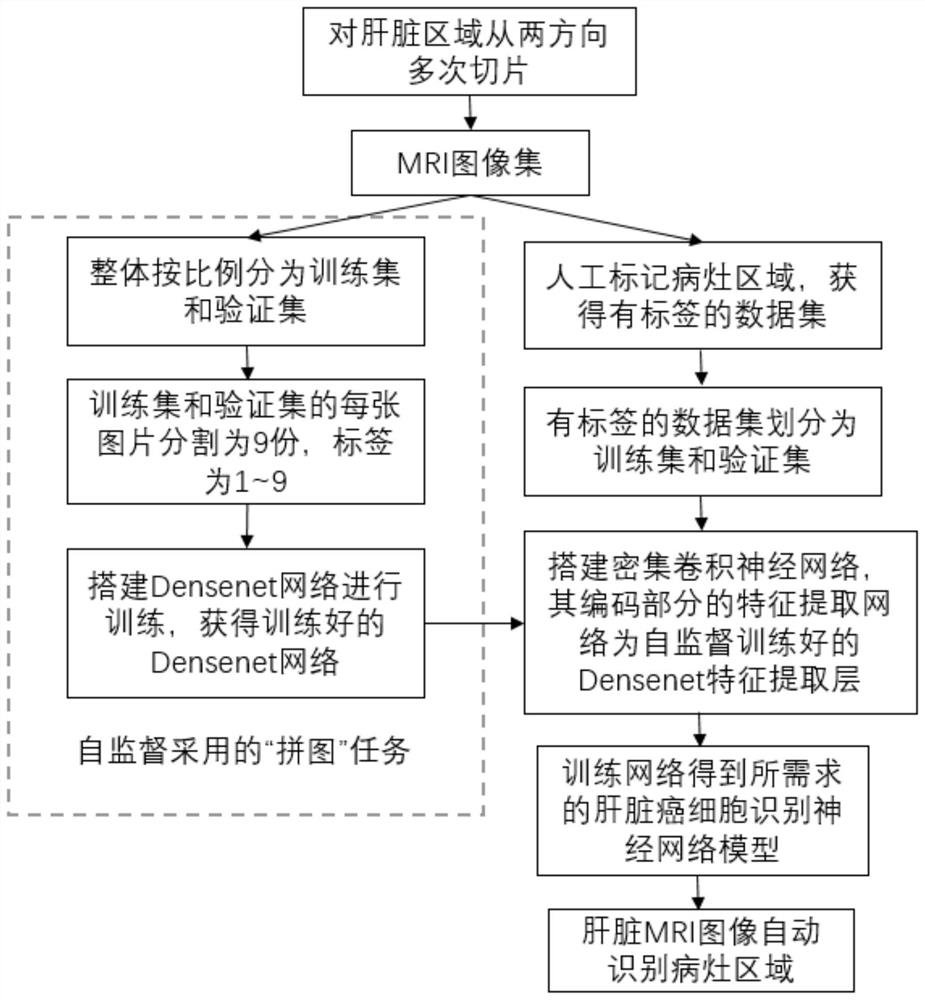
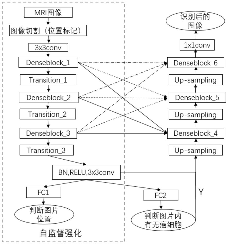
[0039] refer to figure 1 , a liver tumor identification method based on a self-supervised dense convolutional neural network, including the following steps:

[0040] Step 1. According to the patient's magnetic resonance image, slice from two cross directions to obtain the liver slice dataset {A};

[0041] Specifically, a liver magnetic resonance imaging (MRI) technique is used to acquire medical images. Slices are made from two intersecting directions, assuming that the conventional medical slice direction is the main direction to cut M slices, and a direction perpendicular to the main direction is selected as the auxiliary direction to cut N slices, and a total of M+N slices can be obtained from one patient , where M and N are assumed to be 10, and the MRI slice images of multiple pat...


PUM


Abstract

The invention discloses a liver tumor recognition method based on a self-supervised dense convolutional neural network. The method comprises the following steps: obtaining a liver slice data set from a magnetic resonance image of a patient, carrying out the segmentation of the slice data set, training a constructed dense convolutional network, enabling the trained dense convolutional network to serve as a coding module, constructing a self-supervised learning network, and carrying out the recognition of a liver tumor through the self-supervised learning network. Through dense connection of the coding modules, a tumor region of a part of an image in a slice data set is manually marked, then the whole slice data set is segmented, segmented image blocks are adopted to train a self-supervised learning network, and the trained self-supervised learning network is adopted to automatically identify tumors in the image. The dense convolutional neural network based on self-supervision is used for liver tumor recognition, a puzzle task is set as a self-supervision upstream training task, useful representations are learned from a large number of images which are not subjected to medical labeling and are used for learning training of downstream target tasks, and therefore the purposes of automatically expanding training data samples and improving the recognition efficiency are achieved. The dependence on expert experience and historical data is reduced, and the recognition accuracy of the liver lesion region is improved.

Description

technical field [0001] The invention belongs to the technical field of medical image recognition, in particular to a liver tumor recognition method based on a self-supervised dense convolutional neural network. Background technique [0002] Liver cancer (hepatocellular carcinoma, HCC) is one of the malignant tumors with the highest mortality and morbidity in the world. Although surgical resection is the first choice for the treatment of liver cancer, the early symptoms of HCC are usually not obvious in clinical practice. In the middle and advanced stages, or due to conditions such as severe liver cirrhosis and abnormal liver function, less than 25% of clinically discovered HCC can be surgically resected. For patients who do not have the conditions for surgical resection, transarterial chemoembolization (Transarterial chemoembolization, TACE)) is the main choice. The theoretical basis of TACE for the treatment of HCC is that 70%-75% of the blood supply of normal liver tissue...

Claims


Application Information

Patent Timeline
Patent Type & Authority Applications(China)
IPC IPC(8): G06T7/00G06T7/11G06K9/38G06K9/46G06K9/62G06N3/04G06N3/08
CPCG06T7/0012G06T7/11G06N3/08G06T2207/10088G06T2207/20076G06T2207/20081G06T2207/20084G06T2207/30056G06T2207/30096G06N3/047G06N3/048G06N3/045G06F18/2415G06F18/241
Inventor 潘奇李凯旋杜佳忆任芳杨自华李鹏杨延延王梦祥
Owner SECOND AFFILIATED HOSPITAL OF XIAN MEDICAL UNIV
Why Eureka
Try Eureka