Method for observing biological tissue sample

A tissue and living body technology, applied in the field of observation of living tissue samples, can solve the problem of thinning the sample thickness and so on

Pending Publication Date: 2021-10-12
HITACHI HIGH TECH SCI CORP
View PDF1 Cites 0 Cited by
  • Summary
  • Abstract
  • Description
  • Claims
  • Application Information

AI Technical Summary

Problems solved by technology

[0010] However, in the observation method of a biological tissue sample described in Patent Document 1, a skilled technique is required to cut out the biological tissue, and it is difficult to reduce the thickness of the sample for more precise observation of the sample.

Method used


Image

  • Method for observing biological tissue sample
  • Method for observing biological tissue sample
  • Method for observing biological tissue sample

Examples


Embodiment Construction

[0037] Hereinafter, this embodiment will be described in detail with reference to the drawings as appropriate. In the drawings used in the following description, in order to facilitate understanding of the features of the present invention, characteristic parts are sometimes shown enlarged for convenience, and the dimensional ratios and the like of each component may be different from actual ones. The materials, dimensions, and the like illustrated in the following description are examples, and the present invention is not limited thereto, and can be appropriately changed and implemented within the range in which the effects are exhibited.

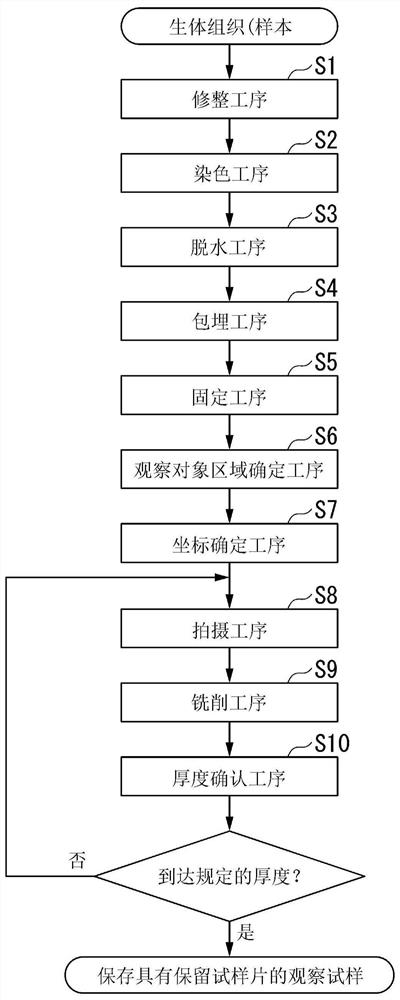
[0038] figure 1 It is a flowchart showing the observation method of a living tissue sample of the present invention step by step.

[0039] As the living tissue applied to the present embodiment, for example, various organs, tissue pieces such as micro-organs produced from iPS cells, cell masses, and the like are exemplified. First, such ...


PUM

PropertyMeasurementUnit
thicknessaaaaaaaaaa
thicknessaaaaaaaaaa

Abstract

Provided is a method for observing a biological tissue sample. As a result, it is possible to easily grasp an observation target region present in a biological tissue, and it is possible to easily cut a plurality of sample pieces from a sample block of the biological tissue. The observation method comprises the following steps: a resin embedding step of forming a sample block; a fixing step in which the sample block is divided into a plurality of sample pieces, and each of the sample pieces is fixed to a sample piece placement member to form a plurality of observation samples; an observation target region specifying step for specifying an observation target region in which precise observation is performed; a coordinate determination step for determining and registering the coordinates of the observation target region on the sample piece for each of the observation samples; a milling step for irradiating the observation target region of the sample sheet with an ion beam or a neutral particle beam using a gas as an ion source with reference to the coordinates, and exposing an observation surface located inside the sample sheet; and an imaging step for acquiring an SEM image of the observation surface using a scanning electron microscope.

Description

technical field [0001] The present invention relates to a method for observing biological tissue samples. Background technique [0002] In the investigation and research of living tissue in the fields of medical biology, regenerative medicine, and pharmaceuticals, it is required to observe the morphology of living tissue three-dimensionally. For example, in regenerative medicine, micro-organs, which are the basis of organs, are fabricated from iPS (induced pluripotent stem cells: induced pluripotent stem cells) cells. Micro-organs are also used in screening for research development in pharmaceuticals. There is a high demand for three-dimensionally grasping the fine structure of organs or tissues by further cutting such micro-organs into several pieces. [0003] However, depending on the capabilities (magnification, depth of focus, and resolution) of microscopes used for observation, stereoscopic observation of living tissue is not necessarily easy, and there are various pr...

Claims


Application Information

Patent Timeline
Patent Type & Authority Applications(China)
IPC IPC(8): G01N1/28G01N1/30G01N1/31G01N1/36G01N21/84G01N23/2251
CPCG01N1/28G01N1/30G01N1/31G01N1/36G01N23/2251G01N21/84G01N2223/6126G01N1/06G01N1/32G01N2001/364G01N1/286G01N2001/2873
Inventor 满欣麻畑达也
Owner HITACHI HIGH TECH SCI CORP
Why Eureka
Try Eureka