Method and device for identifying hyphae in cornea confocal image

A recognition method and confocal technology, applied in the field of medical image processing, can solve problems such as misdiagnosis, similar shape, missed diagnosis, etc.

Pending Publication Date: 2021-11-02
PEKING UNIV THIRD HOSPITAL +1
View PDF5 Cites 1 Cited by
  • Summary
  • Abstract
  • Description
  • Claims
  • Application Information

AI Technical Summary

Problems solved by technology

[0002] The appearance of hyphae in corneal confocal images is an important basis for judging fungal keratitis, but sometimes hyphae and nerve fibers will exist in corneal confocal images at the same time. Both hyphae and nerve fibers are linear structures with similar shapes and very Difficult to distinguish. It requires experienced ophthalmologists to find out the difference between the two and carefully distinguish them. Therefore, different doctors may have different subjective experiences, resulting in missed diagnosis and misdiagnosis
The existing technology does not consider the technology of distinguishing hyphae and nerve fibers, but this is where the difficulty of hyphae identification lies

Method used


Image

  • Method and device for identifying hyphae in cornea confocal image
  • Method and device for identifying hyphae in cornea confocal image
  • Method and device for identifying hyphae in cornea confocal image

Examples


Embodiment Construction

[0020] In order to make the purpose, technical solutions and advantages of the present invention clearer, the technical solutions in the present invention will be clearly and completely described below in conjunction with the accompanying drawings in the present invention. Obviously, the described embodiments are part of the embodiments of the present invention , but not all examples. Based on the embodiments of the present invention, all other embodiments obtained by persons of ordinary skill in the art without creative efforts fall within the protection scope of the present invention.

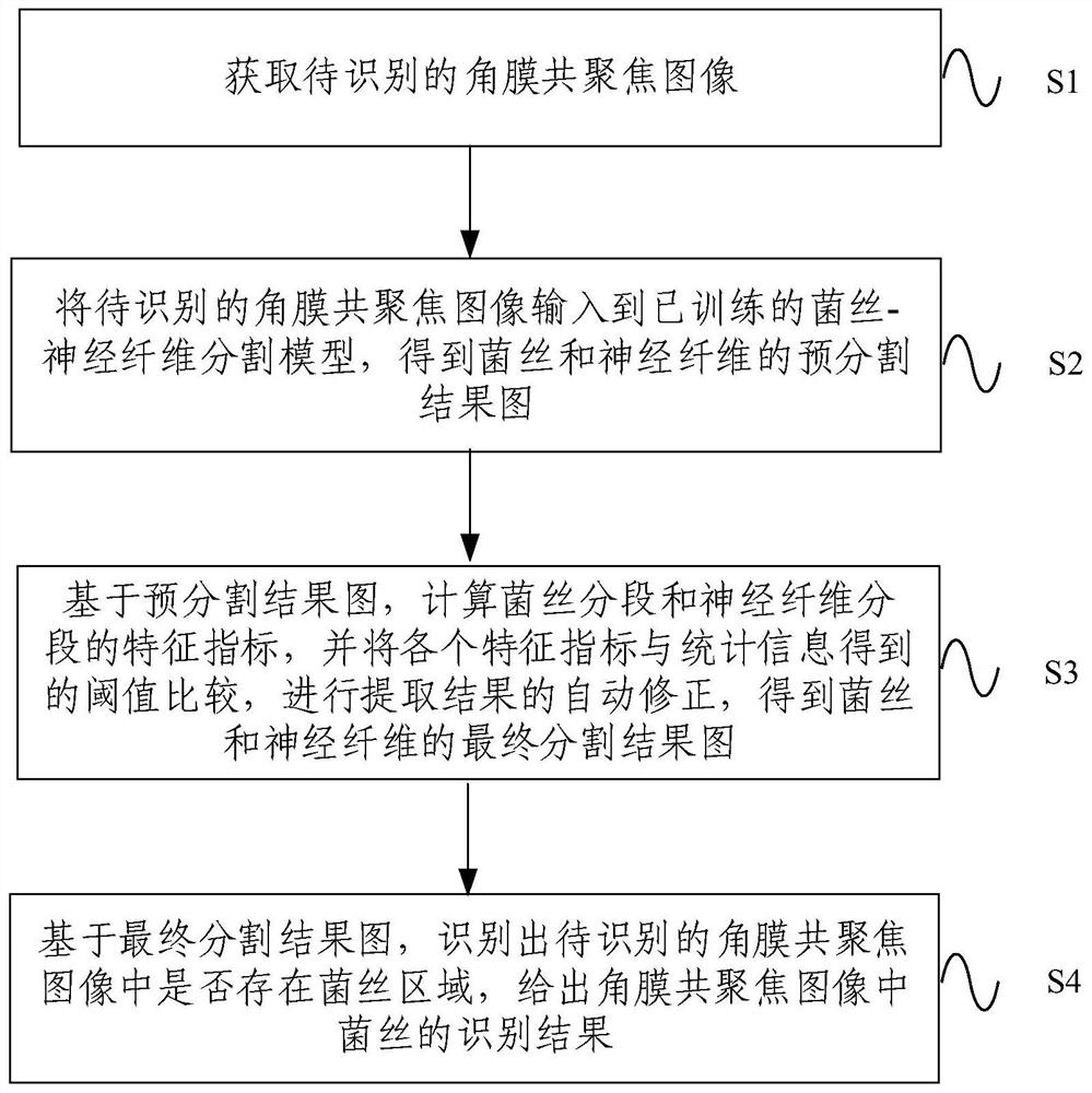
[0021] Combine below Figure 1-Figure 4 The method and device for identifying hyphae in corneal confocal images of the present invention are described.

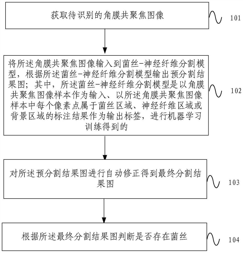
[0022] figure 1 It is one of the flow charts of the hyphae identification method in the corneal confocal image provided by the present invention. Such as figure 1 As shown, the method includes:

[0023] Step 101, acquiring a confocal im...


PUM


Abstract

The invention provides a method and device for identifying hyphae in a cornea confocal image. The method comprises the following steps: acquiring a cornea confocal image to be identified; inputting the cornea confocal image into a hypha-nerve fiber segmentation model, and outputting a pre-segmentation result graph according to the hypha-nerve fiber segmentation model; automatically correcting the pre-segmentation result image to obtain a final segmentation result image; and judging whether hyphae exist or not according to the final segmentation result graph. According to the method and the device for identifying the hypha in the cornea confocal image, the cornea confocal image is input into the hypha-nerve fiber segmentation model, the pre-segmentation result image is output according to the hypha-nerve fiber segmentation model, and the pre-segmentation result image is automatically corrected to obtain the final segmentation result image; and whether hyphae exist or not is judged according to the final segmentation result image, accurate distinguishing of the hyphae and nerve fibers in the image is achieved, and therefore automatic and accurate identification of the hyphae in the cornea confocal image can be achieved.

Description

technical field [0001] The invention relates to the technical field of medical image processing, in particular to a method and device for identifying hyphae in corneal confocal images. Background technique [0002] The appearance of hyphae in corneal confocal images is an important basis for judging fungal keratitis, but sometimes hyphae and nerve fibers will exist at the same time in corneal confocal images. Both hyphae and nerve fibers are linear structures with similar shapes and very It is difficult to distinguish, and it requires experienced ophthalmologists to find out the difference between the two and carefully distinguish them. Therefore, different doctors may have different subjective experiences, resulting in missed diagnosis and misdiagnosis. The prior art does not consider the technology of distinguishing hyphae and nerve fibers, yet this is where the difficulty of hyphae identification lies. Contents of the invention [0003] In order to solve the problems i...

Claims


Application Information

Patent Timeline
Patent Type & Authority Applications(China)
IPC IPC(8): G06K9/00G06K9/62G06N20/00
CPCG06N20/00G06F18/214
Inventor 洪晶秦晓冉彭荣梅程健陈炜
Owner PEKING UNIV THIRD HOSPITAL
Why Eureka
Try Eureka