Image processing method and device

An image processing and image technology, applied in the image field, can solve the problems of affecting segmentation results, low tubular structure recognition rate, noise sensitivity, etc.

Pending Publication Date: 2021-11-26
QINGDAO HISENSE MEDICAL EQUIP
View PDF0 Cites 1 Cited by
  • Summary
  • Abstract
  • Description
  • Claims
  • Application Information

AI Technical Summary

Problems solved by technology

However, these methods have their own defects: the segmentation algorithm based on region growing is very sensitive to noise, the recognition rate is low for tubular structures with small ends and variable anatomical structures, and the segmentation effect is poor; the segmentation accuracy based on level set segmentation algorithm is high , but the excess r

Method used


Image

  • Image processing method and device
  • Image processing method and device
  • Image processing method and device

Examples


Embodiment Construction

[0035] The technical solutions in the embodiments of the present application will be clearly and completely described below in conjunction with the accompanying drawings in the embodiments of the present application. Obviously, the described embodiments are only some of the embodiments of the present application, not all of them. Based on the embodiments in this application, all other embodiments obtained by persons of ordinary skill in the art without making creative efforts belong to the scope of protection of this application.

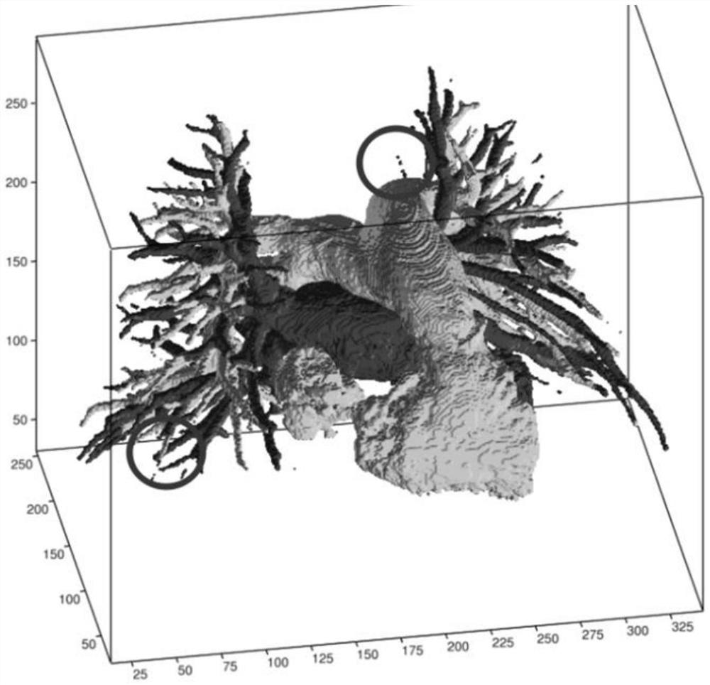
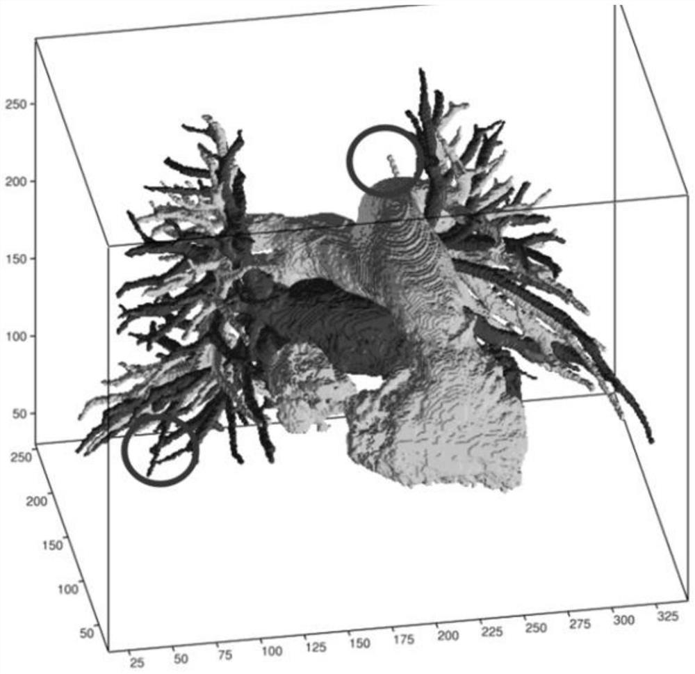
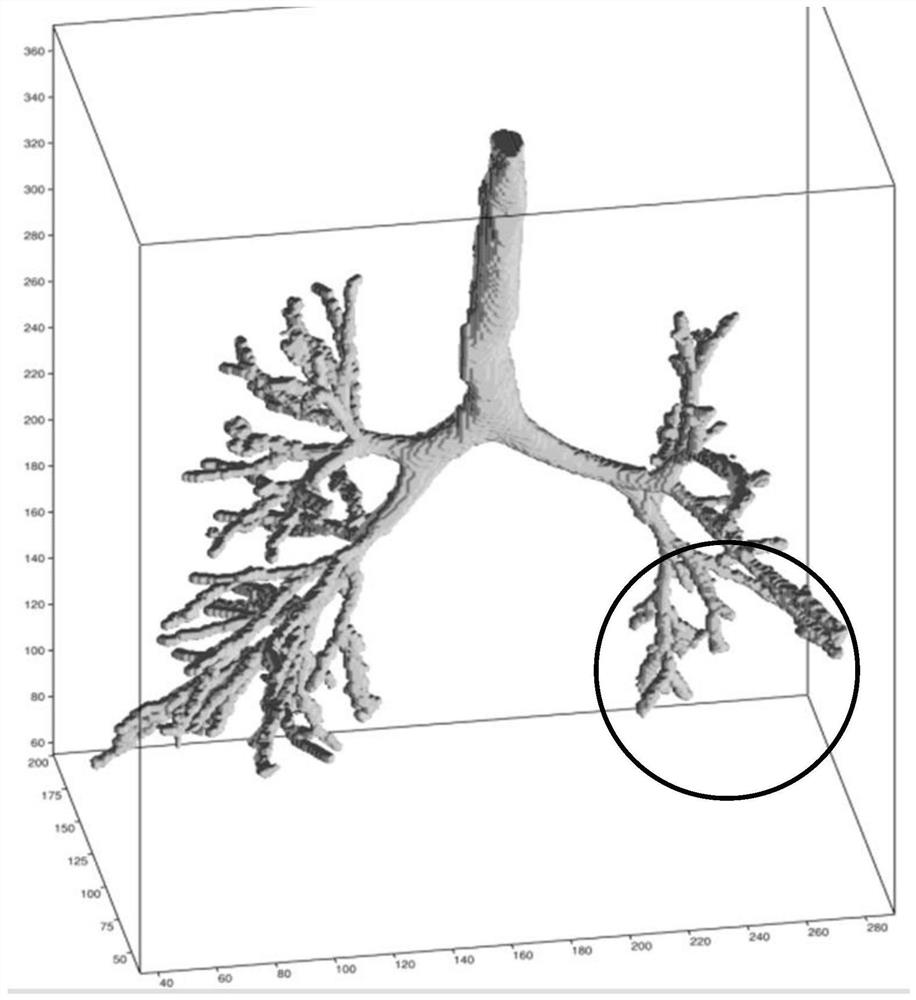
[0036] With the continuous growth of medical needs, the imaging technology of computed tomography (CT) equipment continues to develop, and lung CT examination is currently one of the most commonly performed clinical examinations. Lung CT can accurately display the three-dimensional anatomical structure of lung tissue. After targeted segmentation processing, the three-dimensional structural information of lung parenchyma, pulmonary tracheal tree, and ...


PUM


Abstract

The invention discloses an image processing method and device, which are used for realizing more accurate segmentation of a tiny complex tubular structure in a lung image and obtaining an image which more intuitively displays three-dimensional anatomical structure information of lung parenchyma, a lung tracheal tree and a lung vessel tree. The image processing method provided by the invention comprises the following steps: inputting a to-be-tested sample into a model obtained by training in advance based on a human chest three-dimensional image, and outputting a prediction result, wherein the to-be-tested sample comprises a human chest three-dimensional image of a to-be-tested person; and performing overturning processing on the prediction result to obtain an image including three-dimensional anatomical structure information of the pulmonary parenchyma, the pulmonary tracheal tree and the pulmonary vascular tree.

Description

technical field [0001] The present application relates to the field of image technology, and in particular to an image processing method and device. Background technique [0002] At present, there are relatively few segmentation methods for the tiny and complex tubular structures of the lungs (mainly the pulmonary trachea tree, pulmonary artery tree, and pulmonary vein tree) in computed tomography (CT) images, mainly due to the limitations of previous computer computing power and Today, due to the limitations of manually labeling samples, most of the existing methods are implemented by using traditional segmentation algorithms, mainly including region-based growth, level-set-based and tubular filter enhancement. However, these methods have their own defects: the segmentation algorithm based on region growing is very sensitive to noise, the recognition rate of tubular structures with small ends and variable anatomical structures is low, and the segmentation effect is poor; th...

Claims


Application Information

Patent Timeline
IPC IPC(8): G06T7/10G16H30/20G06N3/08
CPCG06T7/10G16H30/20G06N3/08G06T2207/10081G06T2207/20081G06T2207/20084G06T2207/30204G06T2207/30061
Inventor 郭又文李其花刘于豪田广野陈永健
Owner QINGDAO HISENSE MEDICAL EQUIP
Why Eureka
Try Eureka