Lung tissue dissimilation degree judgment method and device

A technology of lung tissue and degree, applied in the field of judging method and device, method and device for judging the degree of alienation of lung tissue, can solve the problems of weakening HRCT use value, shortage, error, etc., to increase economic burden, without hysteresis , good specific effect

Pending Publication Date: 2020-06-16
NAT UNIV OF DEFENSE TECH
View PDF8 Cites 4 Cited by
  • Summary
  • Abstract
  • Description
  • Claims
  • Application Information

AI Technical Summary

Problems solved by technology

However, this method also has two obvious problems: on the one hand, it cannot directly evaluate the lung dissimilation of the target object, but only indirectly reflects the changes in lung organs or tissues through the difference in functional level, which will lead to a certain lag Generally, the difference is only found when the tissue is alienated enough to seriously affect the function, and it is impossible to accurately observe the patient's lung lesions in real time; on the other hand, this method has a large error, and this method requires the subject to be able to breathe spontaneously And actively cooperate, this method is not suitable for some subjects with poor tolerance, or some patients with spontaneous breathing difficulties, which is also the direct reason for the lack of pulmonary function test results in some patients with acute exacerbation leading to respiratory failure
Although the diagnosis based on HRCT can achieve the accuracy close to that of pathological examination in some cases, at present, HRCT is mainly used to directly observe the imaging features of lung lesions clinically.
There are some guiding principles for the diagnosis of lung lesions based on HRCT, but there is a lack of clear indicators that can be quantitatively analyzed, which leads to differences in the diagnostic opinions of different doctors on CT images, which also weakens the role of HRCT in the diagnosis of lung lesions. use value

Method used


Image

  • Lung tissue dissimilation degree judgment method and device
  • Lung tissue dissimilation degree judgment method and device
  • Lung tissue dissimilation degree judgment method and device

Examples


Embodiment 1

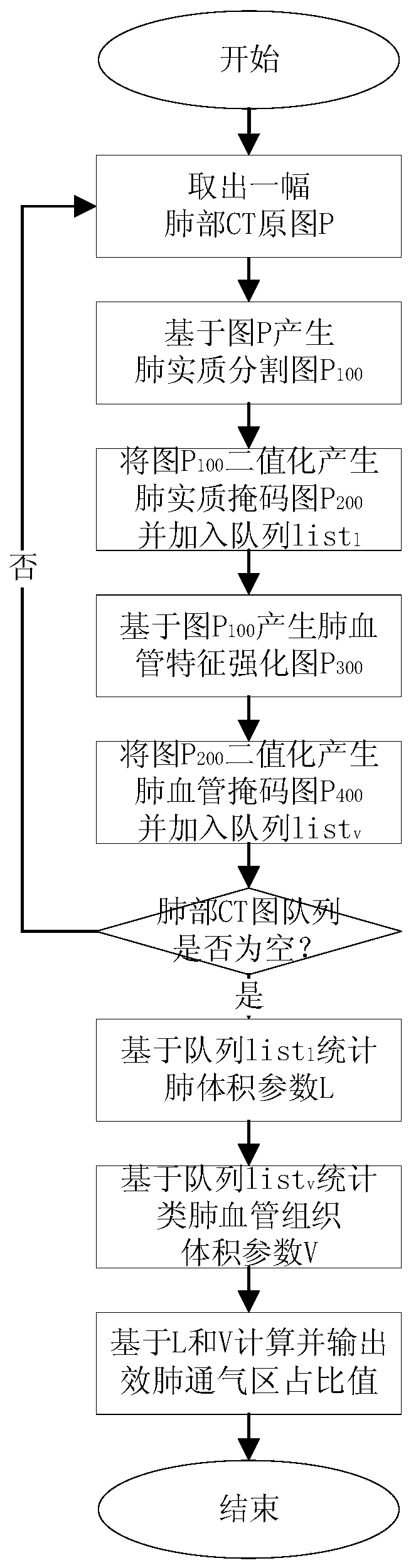
[0170] Three groups of lung CT images were processed. The three groups of samples were from healthy adult men (44 years old), middle-aged women with mild interstitial pneumonia (46 years old), and elderly women with severe interstitial pneumonia (57 years old). ), to determine the degree of lung tissue dissimilation, the method flow is as follows figure 1 shown.

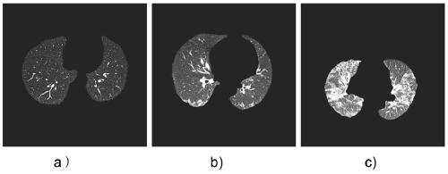
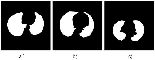
[0171] (1) Take a lung CT image P from the lung CT image queue for lung parenchyma segmentation, and generate a lung parenchyma segmentation map P 100 ,Such as figure 2 as shown ( figure 2 a is a healthy sample image, figure 2 b is the image of mild lung tissue abnormality, figure 2 c is a severe lung tissue alienation image); (2) Set the pixel value of the lung parenchyma region to a uniform non-zero value, and set the background pixel value to 0 to generate a lung parenchyma mask image P 200 ,Such as image 3 as shown ( image 3 a is a healthy sample image, image 3 b is the image of mild lung tissue ab...

Embodiment 2

[0175] Edge segmentation is performed on the CT image of interstitial lung disease. Affected by the disease, the edge of the lung in the CT image is broken and interfered by other organs. There is a hole in the middle of the lung parenchyma in the CT image. The method flow is as follows Image 6 shown.

[0176] (1) Define the CT image of interstitial lung disease as image P, such as Figure 7 (2) Carry out binarization processing on the lung CT image P and perform median filter denoising to form a binarized image P 2 , where the gray value of the pixel in the lung parenchyma region is set to 1, and the gray value of the pixel in the background region is set to 0, such as Figure 8 Shown; (3) Choose a Gaussian convolution kernel with a standard deviation of σ (set to 0.5), for P 2 Perform convolution operation to smooth the edge area of ​​the image to form a graph P 3 ; (4) Use the Laplacian operator to smooth and filter the graph P 3 For processing, select the outer bounda...


PUM


Abstract

The invention provides a lung tissue dissimilation degree judgment method and device, and the method comprises the following steps: carrying out the pulmonary parenchyma segmentation of all lung CT images, and generating a pulmonary parenchyma segmentation image; performing binarization processing on the pulmonary parenchyma segmentation image to generate a pulmonary parenchyma mask image; carrying out feature enhancement processing on pulmonary vessels in all pulmonary parenchyma segmentation images to generate pulmonary vessel feature enhancement images; carrying out binarization processingon the pulmonary vessel feature enhancement image to generate a pulmonary vessel mask image; counting the number L of pulmonary parenchyma region pixel points in all pulmonary parenchyma mask images to serve as volume parameters of the pulmonary parenchyma region pixel points; counting the number V of pulmonary vessel region pixel points in all the pulmonary vessel mask images as volume parametersof the pulmonary vessel region pixel points; and based on the number L of the pulmonary parenchyma region pixel points in all the pulmonary parenchyma mask images and the number V of the pulmonary vessel region pixel points in all the pulmonary vessel mask images, calculating to obtain a pulmonary effective ventilation function region proportion ELVAR value, and outputting the ELVAR value. The method and the device provided by the invention provide a reliable basis for clinical related lung condition evaluation.

Description

technical field [0001] The invention belongs to the field of medical image processing and relates to a disease (the disease can be seen in diseases that cause changes in lung tissue CT images, such as influenza A H1 N1 pneumonia, interstitial pneumonia, 2019-nCov virus pneumonia, severe acute respiratory syndrome Syndrome (SARS) etc.), in particular to a method and device for judging the degree of alienation of lung tissue. Background technique [0002] Pulmonary function test is an important means of clinical examination of respiratory diseases, mainly used for the detection of airway patency and lung volume, for early detection of lung and airway lesions, identification of causes of dyspnea, diagnosis of lesion sites, and serious diseases It has important clinical value in the evaluation of the degree and prognostic effect, the evaluation of the curative effect of drugs or other treatment methods, and the evaluation of the patient's surgical tolerance or labor intensity to...

Claims


Application Information

Patent Timeline
IPC IPC(8): G06T7/00G06T7/11G06T7/136G06T7/181G06T7/62G06T5/00G06T5/20
CPCG06T7/0012G06T7/11G06T7/136G06T7/181G06T7/62G06T5/20G06T2207/10081G06T2207/30061G06T2207/30101G06T2207/20024G06T5/70Y02A90/10
Inventor 汪昌健李方召郭凌超
Owner NAT UNIV OF DEFENSE TECH
Why Eureka
Try Eureka