Pulmonary nodule detection method and system

A detection method, a technology for pulmonary nodules, applied in the field of medical image processing, can solve the problems of low detection accuracy, low automation, and low efficiency of nodule detection, and achieve the elimination of false positive nodules, improvement of segmentation accuracy, and improvement of detection accuracy Effect

Inactive Publication Date: 2020-10-09
南京冠纬健康科技有限公司
View PDF4 Cites 4 Cited by
  • Summary
  • Abstract
  • Description
  • Claims
  • Application Information

AI Technical Summary

Problems solved by technology

However, traditional CAD systems are based on some artificially pre-designed low-level features, such as grayscale, texture, shape
However, the shape, size and texture of real nodules in the lung parenchyma are highly variable, and low-level feature descriptions cannot fully represent these real nodules, so the detection accuracy of nodules is not high
At the same time, traditional CAD systems often have many operation steps, which need to be completed manually, and the flexibility is poor and end-to-end inspection cannot be carried out, so the degree of automation of inspection is low and the efficiency is not high.
[0004] At present, deep learning methods are rarely used in the field of lung parenchyma segmentation. Although there are many traditional methods that can better solve the problem of lung parenchyma segmentation, lung parenchyma segmentation based on deep learning is still a direction worth exploring.

Method used


Image

  • Pulmonary nodule detection method and system
  • Pulmonary nodule detection method and system
  • Pulmonary nodule detection method and system

Examples


Embodiment Construction

[0081] The following will clearly and completely describe the technical solutions in the embodiments of the present invention with reference to the accompanying drawings in the embodiments of the present invention. Obviously, the described embodiments are only some, not all, embodiments of the present invention. Based on the embodiments of the present invention, all other embodiments obtained by persons of ordinary skill in the art without making creative efforts belong to the protection scope of the present invention.

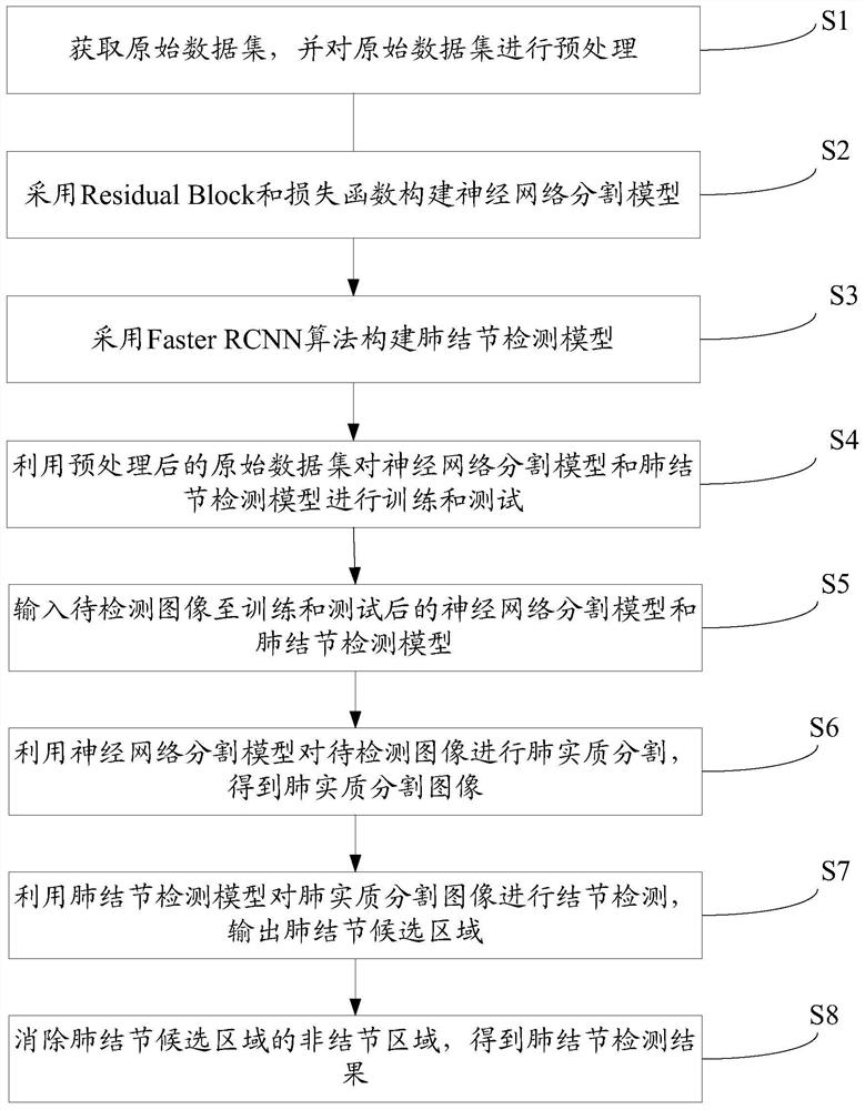
[0082] Such as figure 1 As shown, the embodiment of the present invention discloses a method for detecting pulmonary nodules, comprising the following steps:

[0083] S1. Obtain the original data set and preprocess the original data set;

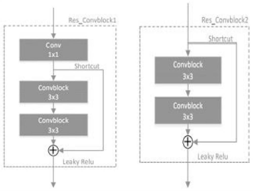
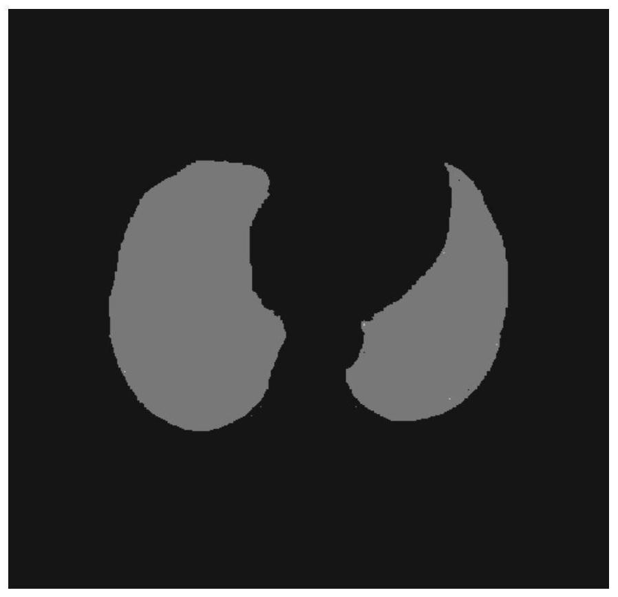
[0084] S2. Construct a neural network segmentation model by using Residual Block and loss function;

[0085] S3, using the Faster RCNN algorithm to construct a pulmonary nodule detection model;

[0086] S4. Using the prep...


PUM


Abstract

The invention discloses a pulmonary nodule detection method which comprises the following steps: acquiring an original data set, and preprocessing the original data set; constructing a neural networksegmentation model by adopting a Residual Block and a loss function; constructing a pulmonary nodule detection model by adopting a Faster RCNN algorithm; training and testing a neural network segmentation model and a pulmonary nodule detection model by using the preprocessed original data set; inputting a to-be-detected image to the trained and tested neural network segmentation model and the pulmonary nodule detection model; performing lung parenchyma segmentation on the to-be-detected image by using the neural network segmentation model to obtain a lung parenchyma segmentation image; performing nodule detection on the pulmonary parenchyma segmentation image by using a pulmonary nodule detection model, and outputting a pulmonary nodule candidate region; and eliminating a non-nodule regionof the pulmonary nodule candidate region to obtain a pulmonary nodule detection result. According to the invention, pulmonary parenchyma segmentation and pulmonary nodule detection are respectively carried out based on the neural network segmentation model and the pulmonary nodule detection model, so that the detection precision and efficiency are greatly improved.

Description

technical field [0001] The present invention relates to the technical field of medical image processing, and more specifically relates to a pulmonary nodule detection method and detection system. Background technique [0002] Cancer has become the number one killer that threatens human health in the 21st century. Because the symptoms of early lung cancer patients are not obvious, it is difficult to be detected. When a patient sees a doctor due to clinical symptoms such as chest pain, cough, or even hemoptysis, lung cancer has often developed into an advanced stage. At this time, the patient has missed the best period for treatment, and the mortality rate will double compared with the early stage of lung cancer. Therefore, early screening of lung cancer is very important. The main manifestation of lung cancer in the early stage is pulmonary nodules. There are many types of pulmonary nodules. [0003] With the development of computer technology, the introduction of Computer ...

Claims


Application Information

Patent Timeline
Patent Type & Authority Applications(China)
IPC IPC(8): G06T7/00G06T7/12G06T7/13G06T7/11G06T7/149
CPCG06T7/0012G06T2207/10081G06T2207/20081G06T2207/20084G06T2207/20116G06T2207/30064G06T7/11G06T7/12G06T7/13G06T7/149
Inventor 吴正德刘倩
Owner 南京冠纬健康科技有限公司
Why Eureka
Try Eureka