Laser doppler perfusion imaging with a plurality of beams

a technology of laser doppler and perfusion imaging, applied in the direction of color/spectral property measurement, indication/recording movement, sensors, etc., can solve the problem that the speed of the system is not enough to resolve the fast changes in the doppler signal, and the scanning takes an appreciable time. problems, to achieve the effect of fast and reliable measurements and analysis

Inactive Publication Date: 2005-08-25
PERIMED
View PDF6 Cites 93 Cited by
  • Summary
  • Abstract
  • Description
  • Claims
  • Application Information

AI Technical Summary

Benefits of technology

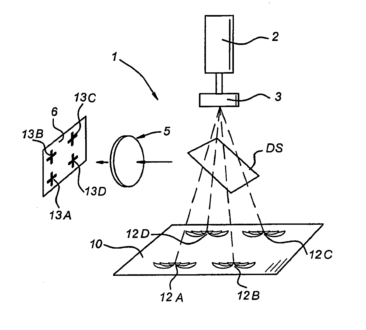
[0021] By using a beam profile comprising a plurality of beams arranged in a pattern, advantageously the Laser Doppler Perfusion Imaging system according to the present invention allows an increase of intensity in the beams, which increases the signal-to-noise-ratio during measurements. Thus, accuracy of detecting a signal is improved. Further, the application of increased intensity by using a plurality of beam profiles allows depth-resolved Laser Doppler Perfusion Imaging measurements.
[0025] Further, due to the characteristics of two-dimensional random access high pixel readout rate image sensors, in a Laser Doppler Perfusion Imaging system according to the present invention faster and more reliable measurements and analysis are possible than in Laser Doppler Perfusion Imaging systems of the prior art.

Problems solved by technology

In both systems the scanning takes an appreciable time since the scanning speed is limited by the trade-off between laser Doppler signal lower cut-off frequency and image quality.
Also, a substantial rise in the noise level at low frequencies appears when using a continuously moving beam as used by Essex et al.
However, the speed of the system was not enough to resolve fast changes in the Doppler signal.
The main disadvantage of LASCA compared with the other techniques is the loss of resolution caused by the need to avenge over a block of pixels in order to produce the spatial statistics used in the analysis.
Main drawbacks of present laser Doppler imagers are: Use of moving parts (mirrors for laser beam steering, or scanning) causes disturbance of the signal.
Mechanical vibration of the scanning mirror deteriorates the measurement accuracy.
Scanning beam 2-D flow imaging takes long time: the time or space resolution of the system is poor.
Present full-frame laser imagers have also either poor time or poor spatial resolution limited by the dynamic range of the detector by Konishi et al or by the imaging approach of Briers et al., respectively, Superimposing the mapped flow onto a digital photograph of the examined area is often difficult since flow-detecting part (photodiode) and observation part (CCD) of the imagers are separated.
Dependence of the signal response on the tissue surface reflectivity affects correct interpretation of measurements on heterogeneous tissue surface: if tissue has a high reflectivity contrast, saturation or under-sensitivity of the photo-detector causes distortion of the flow map.
However, for laser Doppler applications, this kind of detector suffers from two major disadvantages.
Moreover, the dynamic range of the CCD camera is strongly limited by the DC light level inherent in the signal that reaches the detector.
This limitation makes CCD cameras unsuitable for laser Doppler imaging.

Method used


Image

  • Laser doppler perfusion imaging with a plurality of beams
  • Laser doppler perfusion imaging with a plurality of beams
  • Laser doppler perfusion imaging with a plurality of beams

Examples


Embodiment Construction

[0037] As an alternative for detectors applied in prior art Laser Doppler Perfusion Imaging systems, in the present invention a two-dimensional random access high pixel readout rate image sensor such as a CMOS image sensor is used. Those skilled in the art will recognise that any type of MOS-based sensor may be used as detector as well.

[0038] CMOS (Complementary Metal Oxide Semiconductor) image sensors progressively developed over the last few years. CMOS image sensor devices are in some way very similar to CCDs. Both technologies are based on photosensitive diodes or gates in silicon. In both solid state devices photons are converted into an electrical charge. The main difference between CMOS and CCD image sensors is basically, that a CMOS image sensor is a 2-D matrix of photodiodes, which can be addressed randomly at a high sampling rate (as described by B. Dierickx et al., in “Random addressable active pixel image sensors”, Proc. SPIE 2950, 2-7 (1996)), while in CCD sensors the ...


PUM

PropertyMeasurementUnit
distanceaaaaaaaaaa
output poweraaaaaaaaaa
wavelengthaaaaaaaaaa

Abstract

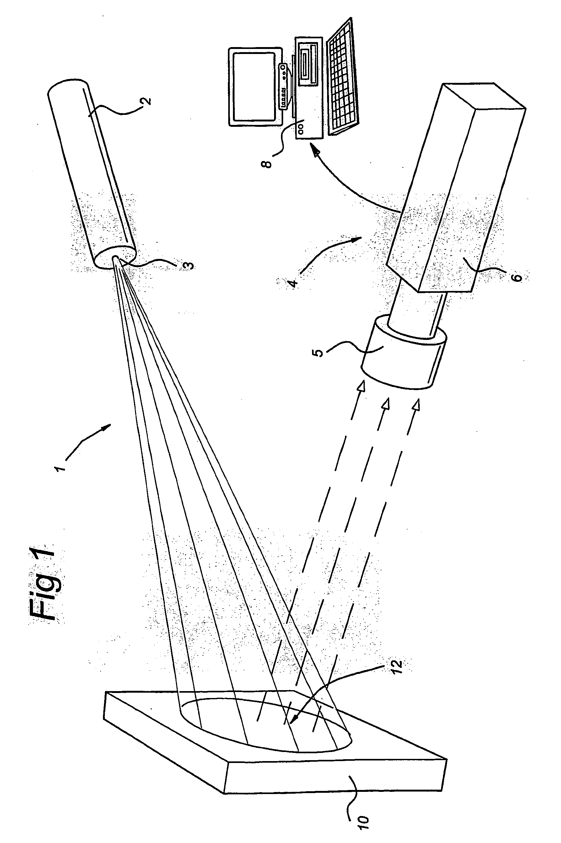
Laser Doppler Perfusion Imaging system including a laser light source (2), an image detector (4), and signal detector (6); the laser light source (2), in use, being arranged for illuminating a selected area of interest (12) on a sample (10) for determining perfusion related data of the sample (10), the image detector, in use, being arranged for capturing an image of the sample (10), and the signal detector (6), in use, being arranged for detecting a laser Doppler signal from the selected area of interest (12) on the sample (10), wherein the laser light source (2) comprises optical means (3) for creating a laser beam comprising a plurality of beams.

Description

FIELD OF THE INVENTION [0001] The present invention relates to a Laser Doppler perfusion Imaging system as defined in the preamble of claim 1. Furthermore, the present invention relates to a method to be carried out in such a system as defined in the preamble of claim 12. PRIOR ART [0002] Laser Doppler Perfusion Imaging (LDPI) is a relatively new non-invasive non-contact optical technique to measure the blood flow in skin and other human tissues. With this technique visualisation of blood flow over a large tissue area is possible. The outcome of the method is a 2-D map of blood perfusion defined as the average blood cell velocity multiplied by the blood cell concentration in the measuring volume. [0003] When tissue is illuminated by laser light, some of the photons are scattered by moving blood cells and undergoes a slight frequency shift due to the Doppler effect. The mixing of the Doppler shifted and zero-Doppler-shifted (unshifted) photons on the remote photodetector causes inten...

Claims


Application Information

Patent Timeline
Patent Type & Authority Applications(United States)
IPC IPC(8): G01P13/00A61B5/026A61B5/0285G01N21/27G01N21/47
CPCA61B5/0261
Inventor SEROV, ALEXANDER NIKOLAEVICHWIENDELT, STEENBERGENDE MUL, FRITS FRANS MARIA
Owner PERIMED
Why Eureka
Try Eureka