
Route planning nose environment modeling method and device

A path planning and modeling method technology, applied in the field of medical image processing, can solve the problems of complex and changeable nasal environment and difficult construction of nasal environment map, and achieve the effect of precise construction

Pending Publication Date: 2021-11-26
THE EYE HOSPITAL OF WENZHOU MEDICAL UNIV +1
View PDF0 Cites 2 Cited by
  • Summary
  • Abstract
  • Description
  • Claims
  • Application Information

AI Technical Summary

Problems solved by technology

However, due to the complex and changeable nasal environment, it is difficult to construct a nasal environment map

Method used


Image

  • Route planning nose environment modeling method and device
  • Route planning nose environment modeling method and device
  • Route planning nose environment modeling method and device

Examples


Embodiment Construction

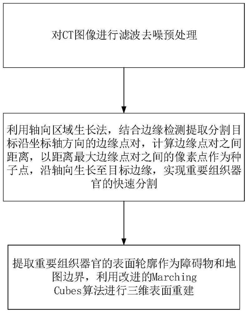
[0016] like figure 1 As shown, this path planning nasal environment modeling method includes the following steps:

[0017] (1) Carry out filter denoising preprocessing to CT image;

[0018] (2) Use the axial region growing method, combined with edge detection to extract the edge point pairs of the segmentation target along the coordinate axis direction, calculate the distance between the edge point pairs, and use the pixel point between the edge point pairs with the largest distance as the seed point, along the axis Growing to the edge of the target to achieve rapid segmentation of important tissues and organs;

[0019] (3) Extract the surface contours of important tissues and organs as obstacles and map boundaries, and use the improved MarchingCubes algorithm for 3D surface reconstruction.

[0020] The present invention preprocesses the CT image by filtering and denoising, uses the axial region growing method, and combines edge detection to extract the edge point pairs of t...


PUM


Abstract

According to the path planning nose environment modeling method and device, accurate construction of a nose environment map can be achieved. The method comprises the following steps: (1) carrying out filtering and denoising preprocessing on a CT image; (2) extracting edge point pairs of a segmentation target along the coordinate axis direction by using an axial region growing method and combining edge detection, calculating the distance between the edge point pairs, and growing to the edge of the target along the axial direction by taking a pixel point between the edge point pair with the maximum distance as a seed point, so as to realize rapid segmentation of important tissues and organs; and (3) extracting surface contours of important tissues and organs as obstacles and map boundaries, and performing three-dimensional surface reconstruction by using an improved Marching Cubes algorithm.

Description

technical field [0001] The invention relates to the technical field of medical image processing, in particular to a path planning nasal environment modeling method and a path planning nasal environment modeling device. Background technique [0002] Important tissues around the nose should be avoided during the surgery. However, due to the complex and changeable nasal environment, it is difficult to construct a nasal environment map. [0003] Different tissues of the human body or animal body have different absorption coefficients for X-rays, and the gray distribution in three-dimensional space obtained after computerized tomography scanning is a CT image. In the CT image space, usually the X-axis is the left-right direction, perpendicular to the X-axis, and the cut plane that divides the image into left and right parts is the sagittal plane; the Y-axis is the front-back direction, perpendicular to the Y-axis, and the cut plane that divides the image into front-back The cut...

Claims


Application Information

Patent Timeline
Patent Type & Authority Applications(China)
IPC IPC(8): G06T17/00G16H50/50G06T3/60G06T5/00G06T7/13
CPCG06T17/00G16H50/50G06T7/13G06T3/608G06T2207/10081G06T5/70
Inventor 杨健吴文灿艾丹妮范敬凡宋红涂云海施节亮
Owner THE EYE HOSPITAL OF WENZHOU MEDICAL UNIV
Learn More