
Cell nucleus segmentation method and device for pathological image

A technology of pathological images and cell nuclei, which is applied in the field of cell nucleus segmentation methods and devices for pathological images, can solve the problems of low accuracy of cell nucleus segmentation results, achieve the effect of reducing learning difficulty and improving training effect

Active Publication Date: 2021-12-07
SHANGHAI BIREN TECH CO LTD
View PDF8 Cites 1 Cited by
  • Summary
  • Abstract
  • Description
  • Claims
  • Application Information

AI Technical Summary

Problems solved by technology

[0004] The present invention provides a cell nucleus segmentation method and device for pathological images to solve the defect of low accuracy of cell nucleus segmentation results in the prior art

Method used


Image

  • Cell nucleus segmentation method and device for pathological image
  • Cell nucleus segmentation method and device for pathological image
  • Cell nucleus segmentation method and device for pathological image

Examples


Embodiment Construction

[0041] In order to make the purpose, technical solutions and advantages of the present invention clearer, the technical solutions in the present invention will be clearly and completely described below in conjunction with the accompanying drawings in the present invention. Obviously, the described embodiments are part of the embodiments of the present invention , but not all examples. Based on the embodiments of the present invention, all other embodiments obtained by persons of ordinary skill in the art without creative efforts fall within the protection scope of the present invention.

[0042] At present, in traditional methods, the gradient algorithm or watershed algorithm is used to segment the nucleus of pathological images. However, for complex pathological images where nuclei are heavily clustered, such as when two tufted nuclei have similar brightness or more tufted nuclei are closely connected, this method cannot accurately identify the outline of the nuclei in the pa...


PUM


Abstract

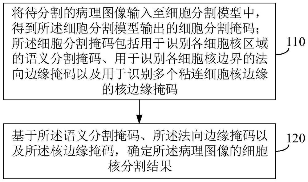
The invention provides a cell nucleus segmentation method and device for a pathological image, and the method comprises the steps: inputting a to-be-segmented pathological image into a cell segmentation model, and obtaining a cell segmentation mask outputted by the cell segmentation model; the cell segmentation mask comprises a semantic segmentation mask used for identifying each cell nucleus region, a normal edge mask used for identifying each cell nucleus boundary and a nucleus edge mask used for identifying a plurality of adhered cell nucleus edges; and determining a cell nucleus segmentation result of the pathological image based on the semantic segmentation mask, the normal edge mask and the nucleus edge mask. The method can greatly improve the precision of the cell nucleus segmentation result.

Description

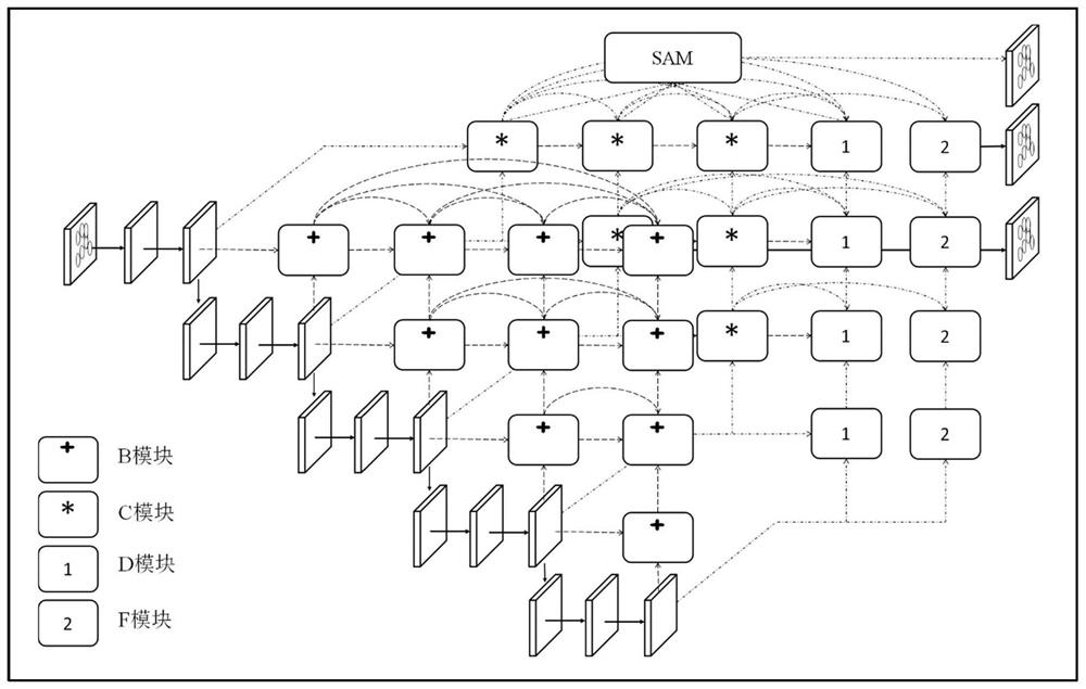
technical field [0001] The present invention relates to the technical field of image processing, in particular to a cell nucleus segmentation method and device for pathological images. Background technique [0002] Cell nucleus segmentation refers to clearly marking the boundary line between each cell and the background or other cells, which is an important basis for microscopic image analysis. [0003] At present, in traditional methods, the gradient algorithm or watershed algorithm is used to segment the nucleus of pathological images. However, for complex pathological images where nuclei are heavily clustered, such as when two tufted nuclei have similar brightness or more tufted nuclei are closely connected, this method cannot accurately identify the outline of the nuclei in the pathological image, that is, the result of the nuclei segmentation Less accurate. Contents of the invention [0004] The present invention provides a cell nucleus segmentation method and devic...

Claims


Application Information

Patent Timeline
Patent Type & Authority Applications(China)
IPC IPC(8): G06T7/00G06T7/11G06T7/12G06T7/13G06T9/20G06K9/62G16H70/60
CPCG06T7/0012G06T7/11G06T7/12G06T7/13G06T9/20G16H70/60G06T2207/10061G06T2207/20081G06F18/214Y02A90/10
Inventor 不公告发明人
Owner SHANGHAI BIREN TECH CO LTD
Learn More