Method for fully automatically measuring lower sternum/xiphoid process in CT/MRI volume

A fully automatic, sternum technology, applied in the field of medical imaging, can solve problems such as affecting the efficiency of medical imaging work, complicated operation methods, etc., to achieve the effect of improving image processing efficiency and processing accuracy, improving work efficiency, and facilitating processing and analysis.

Pending Publication Date: 2022-01-28
CARBON (SHENZHEN) MEDICAL DEVICE CO LTD
View PDF0 Cites 0 Cited by
  • Summary
  • Abstract
  • Description
  • Claims
  • Application Information

AI Technical Summary

Problems solved by technology

However, in the prior art, artificial resolution is usually used to find the lower end of the sternum or the xiphoid process in CT / MRI (magnetic resonance) images, and the operation method is relatively complicated, which seriously affects the work efficiency of medical imaging operations. Therefore, it is necessary to study a A method to automatically measure the lower end of the sternum or xiphoid process in CT / MRI images to solve the above problems

Method used


Image

  • Method for fully automatically measuring lower sternum/xiphoid process in CT/MRI volume
  • Method for fully automatically measuring lower sternum/xiphoid process in CT/MRI volume
  • Method for fully automatically measuring lower sternum/xiphoid process in CT/MRI volume

Examples


Embodiment Construction

[0021] The following will clearly and completely describe the technical solutions in the embodiments of the present invention with reference to the accompanying drawings in the embodiments of the present invention. Obviously, the described embodiments are only some, not all, embodiments of the present invention. Based on the embodiments of the present invention, all other embodiments obtained by persons of ordinary skill in the art without creative work, any modifications, equivalent replacements, improvements, etc., shall be included in the protection scope of the present invention Inside.

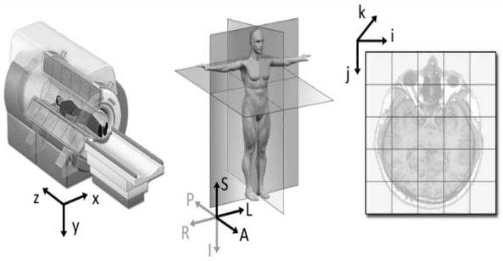
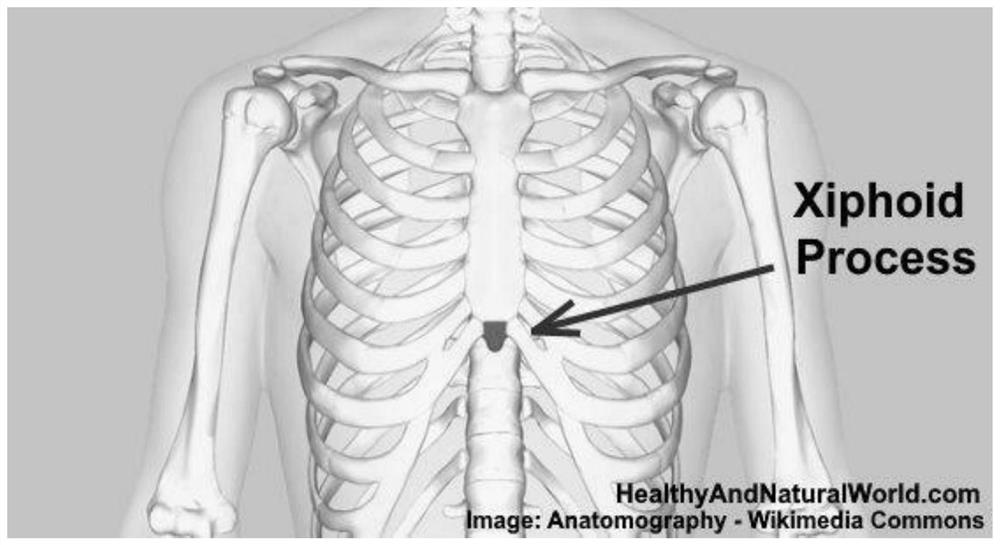
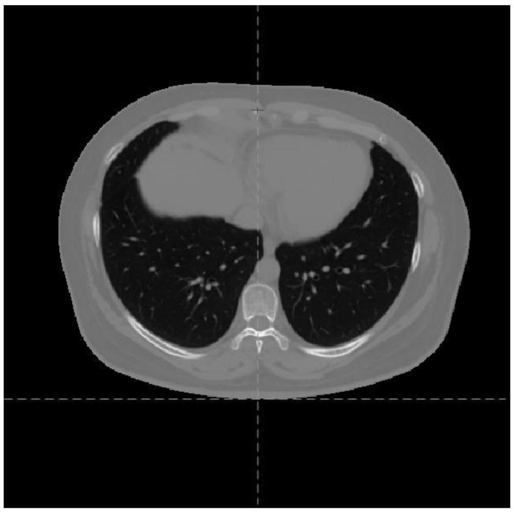
[0022] Such as Figure 1 to Figure 4 As shown, the present embodiment discloses a method for fully automatic measurement of the lower sternal end / xiphoid process in the CT / MRI volume. The search for the lower end of the sternum (SIE) is based on a stack of axial CT / xiphoid processes that conform to the volume scan (the size is MxNxH). Within an MRI slice, a slice-by-slice search is perfo...


PUM


Abstract

The invention discloses a method for fully automatically measuring the lower sternum end/xiphoid process in a CT/MRI volume, and the method comprises the following steps: S1, cutting off a part below a central axis plane in a whole body CT/MRI image, and obtaining an upper body image; S2, cutting off a part, positioned behind a middle coronal plane, in the upper body image to obtain a pre-search image; S3, setting a lower threshold value LT and an upper threshold value UT, and setting a voxel value in the interval [LT, UT] in the pre-searched image as a foreground pixel; S4, establishing a coordinate system and placing the pre-searched image in the coordinate system; S5, searching the slice from the slice at the topmost end; S6, in the current slice, searching a foreground pixel which is located around the median sagittal plane and has the lowest i coordinate; if so, selecting the next level of slice to continue searching; if not, ending the selection; and S7, finally determining the position of the lower end of the sternum or the xiphoid process in the pre-searched image.

Description

technical field [0001] The invention relates to the field of medical imaging, in particular to a method for fully automatic measurement of the lower end of the sternum / xiphoid process in a CT / MRI volume. Background technique [0002] The infrasternal extremity (SIE) or its adjacent xiphoid process (XP) is a bony structure that is easily detected (by simple palpation) in the human thorax and is often used as a reference landmark for different medical applications. However, in the prior art, artificial resolution is usually used to find the lower end of the sternum or the xiphoid process in CT / MRI (magnetic resonance) images, and the operation method is relatively complicated, which seriously affects the work efficiency of medical imaging operations. Therefore, it is necessary to study a A method to automatically measure the lower end of the sternum or xiphoid process in CT / MRI images to solve the above problems. Contents of the invention [0003] The purpose of the present...

Claims


Application Information

Patent Timeline
Patent Type & Authority Applications(China)
IPC IPC(8): G16H30/40
CPCG16H30/40
Inventor 伯斯坦·巴勃罗大卫
Owner CARBON (SHENZHEN) MEDICAL DEVICE CO LTD
Why Eureka
Try Eureka