
Surgical endoscope sheath and urethrobladder endoscope

A technology for endoscope and surgery, which is applied in the field of surgical endoscope sheath and urethral bladder endoscope, and can solve problems such as sphincter injury

Pending Publication Date: 2022-05-31
中国人民解放军总医院第三医学中心
View PDF0 Cites 0 Cited by
  • Summary
  • Abstract
  • Description
  • Claims
  • Application Information

AI Technical Summary

Problems solved by technology

[0003] During the clinical operation, the mirror sheath with the existing structure will put pressure on the sphincter when the diseased tissue is pushed up and down and swings left and right during the operation. When the pressure is too large, the sphincter will be damaged, resulting in urinary incontinence

Method used


Image

  • Surgical endoscope sheath and urethrobladder endoscope
  • Surgical endoscope sheath and urethrobladder endoscope
  • Surgical endoscope sheath and urethrobladder endoscope

Examples


Embodiment Construction

[0023] The following describes in detail the embodiments of the present invention, examples of which are illustrated in the accompanying drawings. The embodiments described below with reference to the accompanying drawings are exemplary, and are intended to explain the present invention and should not be construed as limiting the present invention.

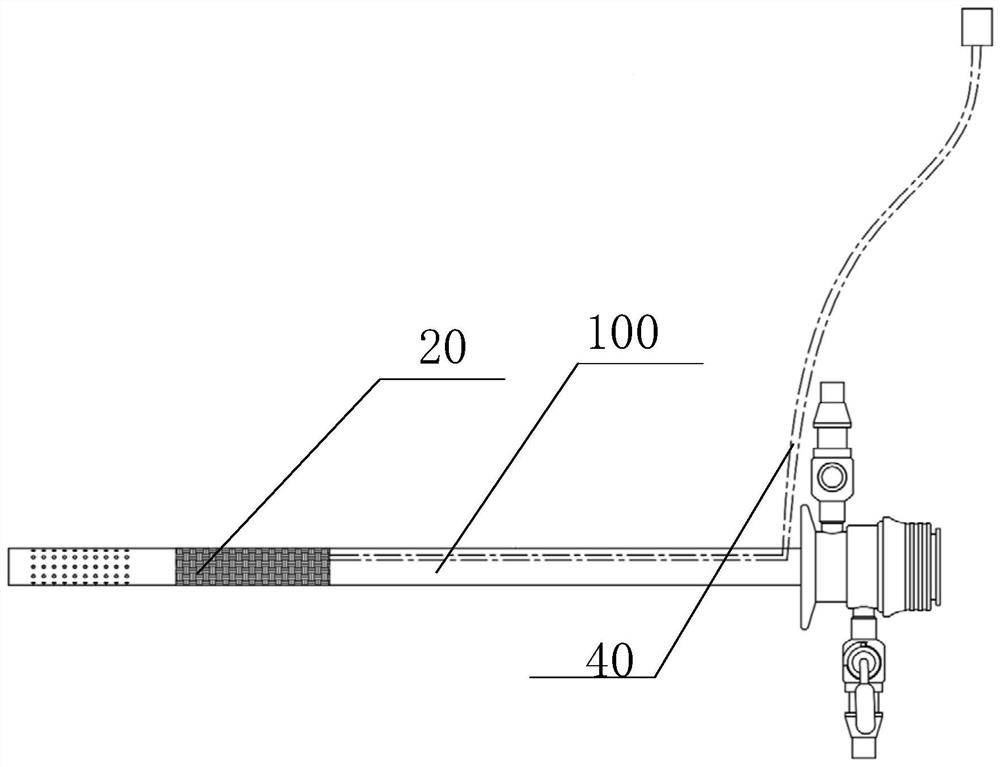
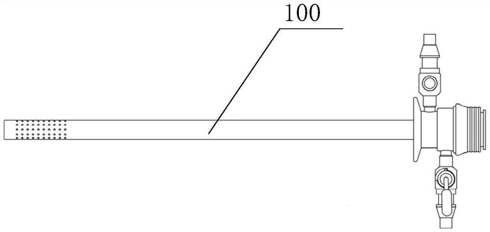
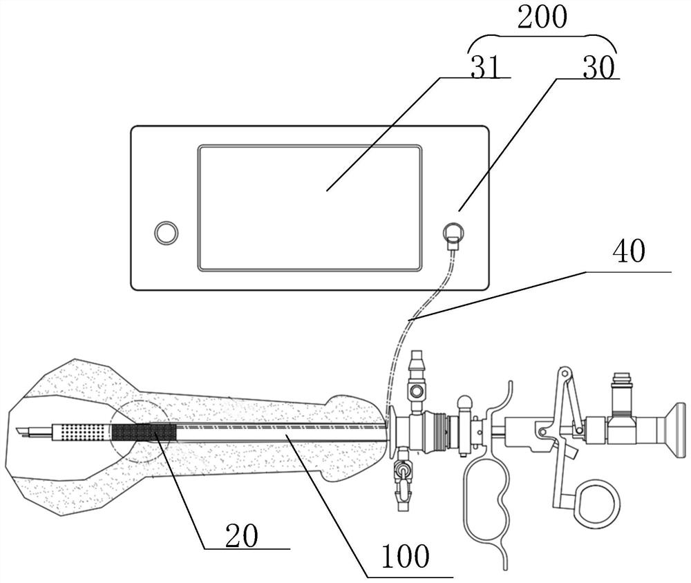
[0024] The surgical endoscope sheath according to the embodiment of the present invention, such as Figure 1-Figure 3 As shown, the surgical endoscope sheath includes a scope sheath tube 100 and a pressure monitoring mechanism 200. The scope sheath tube 100 has a first end and a second end opposite along its length direction, and the first end of the scope sheath tube 100 is provided with a In the quick interface, the pressure monitoring mechanism 200 is connected with the scope sheath tube 100 , and the pressure monitoring mechanism 200 is used to monitor the peripheral wall pressure of the scope sheath tube 100 adjacent to the s...


PUM


Abstract

The surgical endoscope sheath comprises an endoscope sheath tube and a pressure monitoring mechanism, the endoscope sheath tube is provided with a first end and a second end which are opposite to each other in the length direction of the endoscope sheath tube, the first end of the endoscope sheath tube is provided with a quick connector, and the pressure monitoring mechanism is connected with the endoscope sheath tube. The pressure monitoring mechanism is used for monitoring the pressure of the peripheral wall, close to the second end of the endoscope sheath tube, of the endoscope sheath tube. The invention further discloses the urethral bladder endoscope. The surgical endoscope sheath provided by the invention has the advantages that the pressure of sphincter is detected in real time, the sphincter is prevented from being pressed and damaged by the endoscope sheath tube, and the safety is high.

Description

technical field [0001] The invention relates to the technical field of medical equipment, in particular to a surgical endoscope sheath and a urethral cystoscope. Background technique [0002] At present, the sheath of urethral cystoscope in clinical use is mainly composed of a sheath tube, a water inlet valve, a water outlet valve, and a quick interface. When using, first insert the obturator into the urethral cystoscope sheath, insert the endoscopic sheath into the urethra and then pull out the obturator, replace the assembled endoscope and manipulator, and inject normal saline at the inlet valve of the scope sheath, and the water outlet valve. Sewage is released from the place, the user observes the urethra and bladder through the endoscope, and uses the manipulator to perform the operation. During the operation, the endoscope sheath is often required to push the diseased tissue. [0003] In clinical operation, the existing structure of the scope sheath will put pressure ...

Claims


Application Information

Patent Timeline
IPC IPC(8): A61B1/307A61B1/00A61B1/015A61B17/00A61B90/00
CPCA61B1/307A61B1/00131A61B1/00064A61B1/015A61B17/00234A61B90/06A61B90/08A61B2090/065A61B2090/08021A61B2017/00274
Inventor 袁清宋涛陈卫微郝晓伟李宏召张旭
Owner 中国人民解放军总医院第三医学中心
Learn More