Method for segmenting ultrasonic two-dimensional image of thyroid nodule

A thyroid nodule and two-dimensional image technology, applied in the medical field, can solve problems such as general accuracy performance, insufficient accuracy of nodule segmentation, and poor segmentation of edge details, so as to achieve the effect of enhancing the segmentation ability

Pending Publication Date: 2022-07-22
BEIJING TIANTAN HOSPITAL AFFILIATED TO CAPITAL MEDICAL UNIV
View PDF0 Cites 3 Cited by
  • Summary
  • Abstract
  • Description
  • Claims
  • Application Information

AI Technical Summary

Problems solved by technology

However, due to the influence of ultrasound resolution and noise, the precise segmentation of nodules in thyroid ultrasound images is not accurate enough, which easily leads to misjudgment
[0004] The existing thyroid ultrasound image segmentation technology is not good in terms of accuracy, especially in the segmentation of edge details.

Method used


Image

  • Method for segmenting ultrasonic two-dimensional image of thyroid nodule
  • Method for segmenting ultrasonic two-dimensional image of thyroid nodule
  • Method for segmenting ultrasonic two-dimensional image of thyroid nodule

Examples


Embodiment Construction

[0018] In order to make the above objects, features and advantages of the present application more clearly understood, the specific embodiments of the present application will be described in detail below with reference to the accompanying drawings. In the following description, numerous specific details are set forth in order to provide a thorough understanding of the present application. However, the present application can be implemented in many other ways different from those described herein, and those skilled in the art can make similar improvements without departing from the connotation of the present application. Therefore, the present application is not limited by the specific embodiments disclosed below.

[0019] In order to keep the drawings concise, the drawings only schematically show the parts related to the present invention, and they do not represent its actual structure as a product. In addition, in order to make the drawings concise and easy to understand, in...


PUM


Abstract

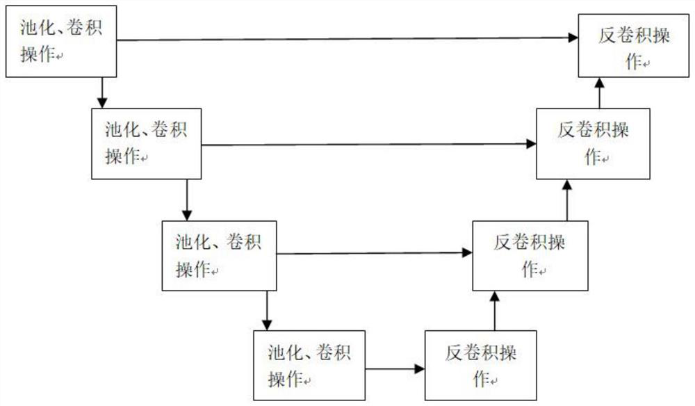
The invention discloses a segmentation method for an ultrasonic two-dimensional image of a thyroid nodule. The segmentation method comprises the following steps: carrying out an image enhancement means on the ultrasonic two-dimensional image; the processed two-dimensional image is input into a model based on a U-net structure, the U-net structure comprises n down-sampling operations and n up-sampling operations, the down-sampling operations are composed of convolution modules of a plurality of different convolution kernels, each convolution module is composed of a pooling operation and a convolution operation, and each convolution operation is composed of a pooling operation and a convolution operation; in the convolution operation, convolution kernels with corresponding sizes are adopted to process an input image, and IN operation and Relu operation are added behind the input image; the up-sampling operation consists of a multi-head self-attention mechanism module and a deconvolution module, the deconvolution module consists of convolution with different convolution kernel sizes and bilinear interpolation or transpose convolution, and IN operation and Relu operation are performed after each convolution operation; a segmentation result is enhanced through a loss function and evaluation is carried out; and outputting the segmented ultrasonic two-dimensional image.

Description

technical field [0001] The present application relates to the field of medical technology, in particular to a method for segmenting an ultrasonic two-dimensional image of a thyroid nodule. Background technique [0002] At present, the number of patients with thyroid cancer is increasing, and the diagnosis and treatment of thyroid cancer are becoming more and more important. At this stage, ultrasound has become an important means of thyroid cancer screening due to its advantages of timeliness and no radiation. However, ultrasound examination is very dependent on the physician's technique and experience, and ultrasound imaging is also easily affected by various factors. In recent years, artificial intelligence technology has developed rapidly, and the use of artificial intelligence to analyze ultrasound images to assist physicians in diagnosing thyroid nodules has also received attention, but how to accurately segment the nodule area is still a relative difficulty. [0003] ...

Claims


Application Information

Patent Timeline
Patent Type & Authority Applications(China)
IPC IPC(8): G06T7/12G06T7/13G06T7/00
CPCG06T7/13G06T7/12G06T7/0012G06T2207/10132G06T2207/20081G06T2207/30096
Inventor 广旸骆伟甘从贵吴佳俊
Owner BEIJING TIANTAN HOSPITAL AFFILIATED TO CAPITAL MEDICAL UNIV
Why Eureka
Try Eureka