Liver medical image registration and fusion method and system

A medical image and fusion method technology, applied in the field of liver medical image processing method and system, can solve the problem of inability to process CT images, achieve multi-directional selectivity and translation without deformation, clear fusion images, demonstration teaching and career growth Helping effect

Pending Publication Date: 2022-08-05
THE AFFILIATED HOSPITAL OF QINGDAO UNIV
View PDF2 Cites 0 Cited by
  • Summary
  • Abstract
  • Description
  • Claims
  • Application Information

AI Technical Summary

Problems solved by technology

However, the image fusion method disclosed in this patent application is mainly used for th

Method used


Image

  • Liver medical image registration and fusion method and system
  • Liver medical image registration and fusion method and system
  • Liver medical image registration and fusion method and system

Examples


Embodiment Construction

[0060] The following describes in detail the embodiments of the present invention, examples of which are illustrated in the accompanying drawings, wherein the same or similar reference numerals refer to the same or similar elements or elements having the same or similar functions throughout. The embodiments described below with reference to the accompanying drawings are exemplary, and are intended to explain the present invention and should not be construed as limiting the present invention.

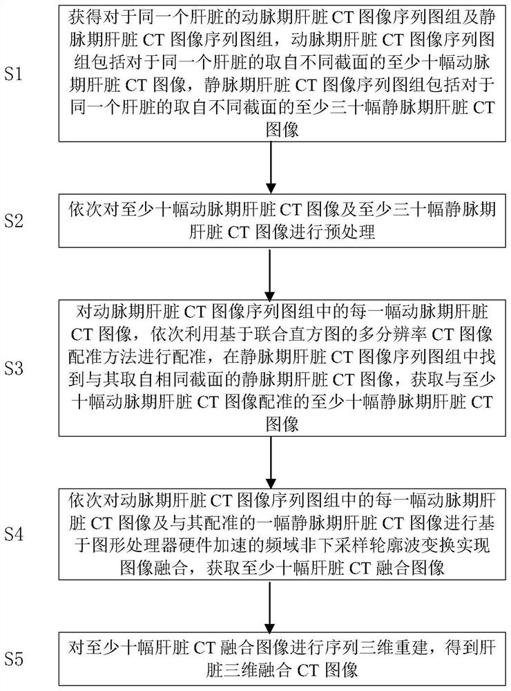
[0061] First, please refer to figure 1 , as a non-limiting embodiment, in the liver medical image registration and fusion method of the present invention, first in step S1, obtain the arterial phase liver CT image sequence map group and the venous phase liver CT image sequence map for the same liver Group, the arterial phase liver CT image sequence map group includes at least ten arterial phase liver CT images taken from different sections of the same liver, and the venous phase liver CT...


PUM


Abstract

The invention discloses a liver medical image registration and fusion method and system, and the method comprises the steps: obtaining an arterial-phase and venous-phase liver CT image sequence chart group for the same liver, and enabling the arterial-phase liver CT image sequence chart group to comprise at least ten arterial-phase liver CT images, the vein phase liver CT image sequence group comprises at least thirty vein phase liver CT images; sequentially registering each arterial phase liver CT image, finding out a vein phase liver CT image with the same cross section from the vein phase liver CT image sequence group, and acquiring at least ten vein phase liver CT images registered with the at least ten arterial phase liver CT images; sequentially fusing each artery-phase liver CT image and one vein-phase liver CT image registered with the artery-phase liver CT image to obtain at least ten liver CT fusion images; and performing sequence three-dimensional reconstruction on the at least ten liver CT fusion images to obtain a liver three-dimensional fusion CT image.

Description

technical field [0001] The present invention relates to the technical field of medical image processing, in particular to a method and system for processing liver medical images. Background technique [0002] The liver is an important organ in the human body. There are many complex piping systems in the liver, such as hepatic artery, hepatic vein, portal vein, and bile duct system. Image processing, visualization research, and biological modeling are of great significance. [0003] The visualization study of the liver can be applied to clinical diagnosis, surgery and liver transplantation. It can help doctors to understand the distribution and connection of the piping system in the liver before surgery, and provide assistance for undiagnosed positioning and surgery. [0004] Medical image registration refers to seeking one or a series of spatial transformations for a medical image to make it spatially consistent with the corresponding points on another medical image. This a...

Claims


Application Information

Patent Timeline
IPC IPC(8): G06T7/00G06T7/13G06T7/136G06T7/37G06T7/38G06T15/10G06T5/40G06T5/00
CPCG06T7/0012G06T7/37G06T7/38G06T7/136G06T7/13G06T15/10G06T5/002G06T5/40G06T2207/10081G06T2207/30101G06T2207/20056G06T2207/30056G06T2207/20192G06T2207/20221
Inventor 魏宾董蒨朱呈瞻董冰子
Owner THE AFFILIATED HOSPITAL OF QINGDAO UNIV
Why Eureka
Try Eureka