Pelvic bone tumor automatic segmentation and three-dimensional reconstruction method based on multi-modal image

A 3D reconstruction and automatic segmentation technology, applied in the field of automatic segmentation and 3D reconstruction of pelvic bone tumors, can solve problems such as loss of limb movement function, large tumor mass, difficulty in bone reconstruction, etc., and achieve reduced segmentation errors, tumor smoothing, and simplified segmentation. the effect of the task

Pending Publication Date: 2022-08-09
DALIAN UNIV OF TECH
View PDF0 Cites 0 Cited by
  • Summary
  • Abstract
  • Description
  • Claims
  • Application Information

AI Technical Summary

Problems solved by technology

Bone tumor is one of the common serious diseases in orthopedics, and the pelvis is one of the predilection sites for bone tumors. Since the early symptoms are not obvious, pelvic tumors are already in the middle and late stages when they are discovered, with large tumor masses and unclear boundaries with surrounding tissues. Coupled with the complex physiological structure of the pelvis, pelvic bone tumors have caused great difficulties for doctors in diagnosis and preoperative planning.
In the treatment of bone tumors, insufficient surgical margin resection will directly lead to the failure of the treatment of tumor lesions; on the contrary, blindly expanding the tumor resection range in order to avoid insufficient tumor resection margins will bring difficulties to bone reconstruction and lead to postoperative loss of limb motor function
In addition, in the repair of bone defects, traditional surgery is based on common experience, ignoring the individual differences in human body structure, which may easily lead to the loss of the mechanical properties of the original bone after repair

Method used


Image

  • Pelvic bone tumor automatic segmentation and three-dimensional reconstruction method based on multi-modal image
  • Pelvic bone tumor automatic segmentation and three-dimensional reconstruction method based on multi-modal image
  • Pelvic bone tumor automatic segmentation and three-dimensional reconstruction method based on multi-modal image

Examples


Embodiment Construction

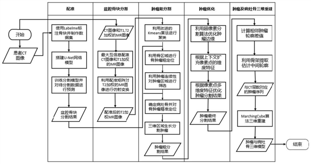
[0062] In order to make the technical solutions and advantages of the present invention clearer, the technical solutions in the embodiments of the present invention will be described clearly and completely below with reference to the accompanying drawings in the embodiments of the present invention:

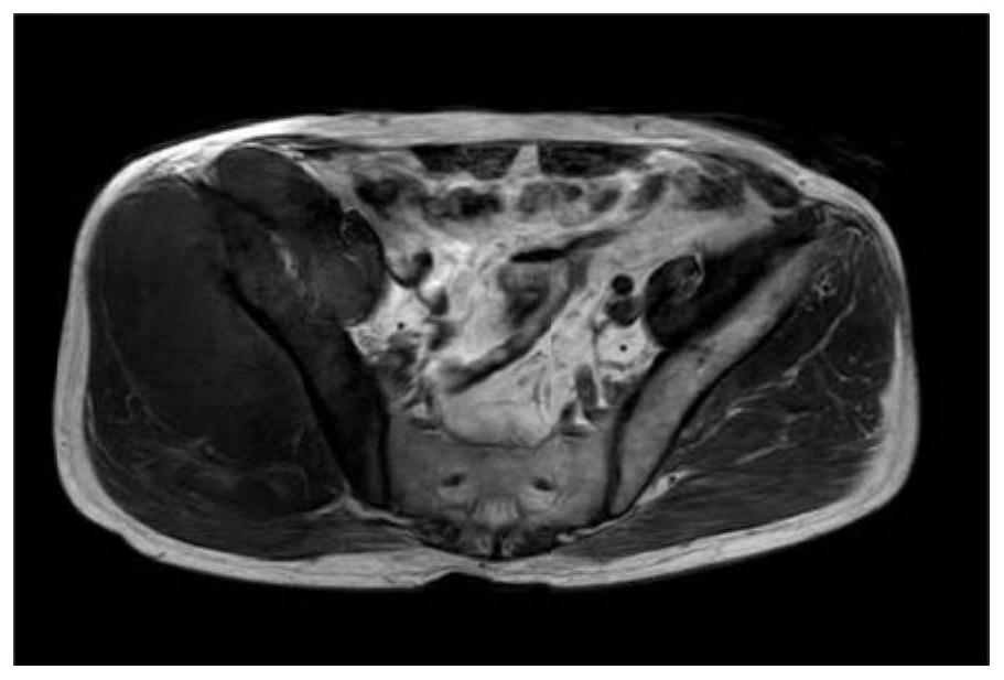
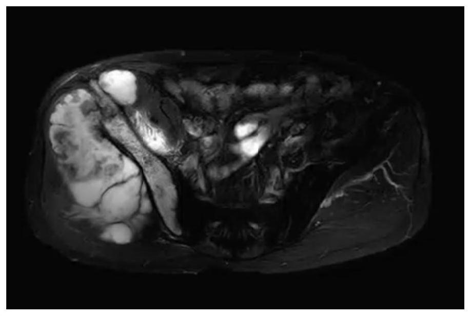
[0063] like figure 1 A method for automatic segmentation and 3D reconstruction of pelvic bone tumors based on multimodal images is shown. In the implementation process, the U-Net network model is used to segment the bone tissue into the ilium, femur and sacrum in the CT image of the patient. , the result is image 3 The three-dimensional reconstruction of the isolated images of the ilium, femur and sacrum was carried out, and the results are as follows Figure 4-6 The position of the sacrum is used to divide the bone tissue into five bone fragments: left ilium, right ilium, sacrum, left femur, and right femur. Then, the mutual information method is used to register the MR image...


PUM


Abstract

The invention discloses an automatic segmentation and three-dimensional reconstruction method for a pelvic bone tumor based on a multi-modal image, and the method achieves the automatic segmentation of an ilium, a sacrum and a femur in a pelvic CT image of a patient through a U-Net network model, and has the advantages that the segmentation error of bone tissues is greatly reduced; secondly, registering the MR image and the CT image by adopting a mutual information method, so that the information of the MR image and the CT image can be comprehensively utilized, and the segmentation task is simplified; and then clustering is carried out on the MR images by adopting an improved K-Means method to obtain candidate regions of tumors, and the method has the advantages that tumor regions can be accurately segmented for MR images with different gray features, so that the method has certain robustness; the method comprises the following steps: screening tumor candidate region images by using a bone region and continuity detection, and effectively solving the problem that a tumor region and a non-tumor region with similar characteristics cannot be distinguished by using medical priori knowledge in the step; in addition, according to the method, the tumor sequence is filled, so that the tumor is smoother during three-dimensional reconstruction and is closer to the real tumor form.

Description

technical field [0001] The invention relates to the field of medical image processing, in particular to a method for automatic segmentation and three-dimensional reconstruction of pelvic bone tumors based on multimodal images. Background technique [0002] With economic development and social progress, human beings urgently need safer, faster, more accurate and more humanized medical services. Therefore, it has become an important scientific research direction to fully pay attention to the individual differences of patients, and combine advanced graphics and image processing technology to carry out personalized and precise surgical design. Bone tumor is one of the common serious diseases in orthopaedics, and the pelvis is one of the most common sites of bone tumors. Due to the insignificant early symptoms, pelvic tumors are already in the middle and late stages when they are discovered, with large tumor masses and unclear boundaries with surrounding tissues. Coupled with th...

Claims


Application Information

Patent Timeline
IPC IPC(8): G06T7/11G06T7/136G06T7/13G06T17/00G06T7/00G06T7/73G06T7/33G06V10/762G06V10/26G06V10/764G06V10/82G06K9/62G06N3/04G06N3/08
CPCG06T7/11G06T7/136G06T7/13G06T17/00G06T7/0012G06T7/73G06T7/33G06V10/763G06V10/267G06V10/764G06V10/82G06N3/08G06T2207/10081G06T2207/20081G06T2207/20112G06T2207/30008G06T2207/30096G06T2207/20084G06N3/047G06N3/048G06F18/23213G06F18/2415
Inventor 刘斌葛琪
Owner DALIAN UNIV OF TECH
Why Eureka
Try Eureka