Method and device for segmenting endocardium and/or epimyocardium of cardiac left ventricle

An epimyocardium and left ventricle technology, applied in the field of image processing, can solve the problems of demanding segmentation accuracy, segmentation deviation, low robustness, etc., to reduce segmentation complexity, reduce segmentation errors, improve segmentation accuracy and The effect of robustness

Active Publication Date: 2022-06-21
SHENZHEN KEYA MEDICAL TECH CORP
View PDF10 Cites 0 Cited by
  • Summary
  • Abstract
  • Description
  • Claims
  • Application Information

AI Technical Summary

Problems solved by technology

Due to the thin-layer structure of the left ventricular myocardium, the direct prediction of the myocardium is very demanding on the accuracy of the segmentation. Without the prior knowledge of other positions as an auxiliary, the problem of segmentation deviation is easy to occur. Therefore, this segmentation idea is in a certain less robust in the sense

Method used


Image

  • Method and device for segmenting endocardium and/or epimyocardium of cardiac left ventricle
  • Method and device for segmenting endocardium and/or epimyocardium of cardiac left ventricle
  • Method and device for segmenting endocardium and/or epimyocardium of cardiac left ventricle

Examples


Embodiment Construction

[0019] In order to make those skilled in the art better understand the technical solutions of the present disclosure, the present disclosure will be described in detail below with reference to the accompanying drawings and specific embodiments. The embodiments of the present disclosure will be described in further detail below with reference to the accompanying drawings and specific embodiments, but are not intended to limit the present disclosure. The steps described herein, if there is no need for a contextual relationship with each other, the order in which they are described herein as an example should not be regarded as a limitation, and those skilled in the art should know that the order can be adjusted as long as It is enough not to destroy the logic between them and make the whole process impossible.

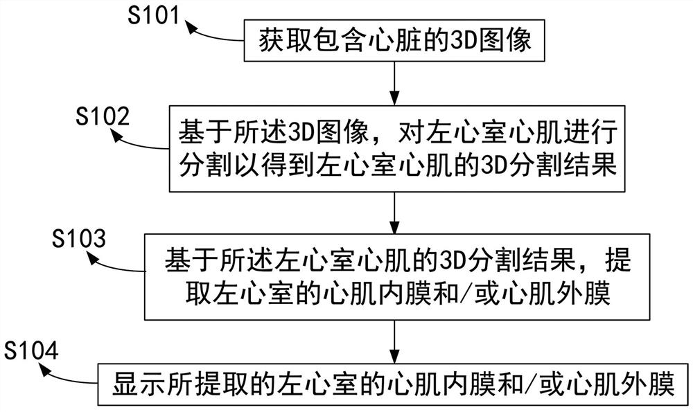
[0020] figure 1 A flow chart illustrating a method of segmenting the endomyocardium and / or epimyocardium of the left ventricle of the heart, which performs the followin...


PUM


Abstract

The present disclosure provides a method for segmenting the endocardium and / or epimyocardium of the left ventricle of the heart, including, via a processor: acquiring a 3D image containing the heart; based on the 3D image, segmenting the left ventricular myocardium to obtain A 3D segmentation result of the left ventricular myocardium; based on the 3D segmentation result of the left ventricular myocardium, extracting the endocardium and / or epimyocardium of the left ventricle; and displaying the extracted endocardium and / or epicocardium of the left ventricle . This method can improve segmentation efficiency, reduce segmentation error, and improve segmentation accuracy.

Description

technical field [0001] The present disclosure relates to the field of image processing, and more particularly, to a method and apparatus for segmenting the endomyocardium and / or epimyocardium of the left ventricle of the heart. Background technique [0002] The segmentation of the left ventricular endocardium and endocardium of the heart is an important step in the analysis of cardiac function. The functional evaluation of the left ventricular myocardial wall thickness and ejection fraction, and the diagnosis and treatment of hypertrophic cardiomyopathy are all based on the accurate segmentation of the left ventricular myocardium and endocardium. In the image of the left ventricle of the heart, because the gray values ​​of the left ventricle endocardium, papillary muscles, trabeculae and other tissues are very close, and the gray value difference between the left ventricle epimyocardium and the external adjacent tissues is small, the segmentation task has a great effect. cha...

Claims


Application Information

Patent Timeline
Patent Type & Authority Patents(China)
IPC IPC(8): G06T7/00G06T7/11G06T7/12G06T7/13G06N3/04G06N3/08
CPCG06T7/0012G06T7/11G06T7/12G06T7/13G06N3/084G06T2207/20081G06T2207/20084G06T2207/30048G06N3/045
Inventor 安木军李俊环丁志敏李育威曹坤琳王立伟
Owner SHENZHEN KEYA MEDICAL TECH CORP
Why Eureka
Try Eureka