Three-dimensional dividing method for medical images

A medical image and three-dimensional technology, applied in the field of medical imaging, can solve the problems of suppressing noise and precise edge positioning at the same time, and achieve fast and accurate interactive segmentation and high application value

Inactive Publication Date: 2007-02-21
PHILIPS & NEUSOFT MEDICAL SYST
View PDF5 Cites 1 Cited by
  • Summary
  • Abstract
  • Description
  • Claims
  • Application Information

AI Technical Summary

Problems solved by technology

That is to say, noise suppression and edge precise positioning cannot be satisfied at the same time.

Method used


Image

  • Three-dimensional dividing method for medical images
  • Three-dimensional dividing method for medical images
  • Three-dimensional dividing method for medical images

Examples


Embodiment Construction

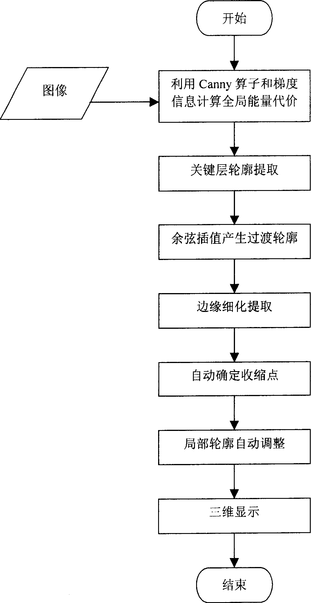
[0043] Combined with the accompanying drawings, such as figure 1The flow chart of the three-dimensional segmentation method of the shown medical image, the detailed segmentation method of the present invention comprises the following three steps:

[0044] (1) Use the canny operator and the gradient field to calculate the energy cost field at the start layer and the end layer, and perform interactive segmentation;

[0045] (2) Cosine interpolation is carried out between the start layer and the end layer, and initial contours are generated on each layer;

[0046] (3) Automatically determine the seed point according to the initial contour between the initial layer and the final layer, and determine the final complete contour through automatic adjustment of the local contour.

[0047] The concrete realization process of the inventive method is as follows:

[0048] step one):

[0049] l(p,q)=w Z f Z (q)+w D f D (p,q)+w G f G (q) (2)

[0050] where f Z (q), f G (q), f ...


PUM


Abstract

A three dimensional method for dividing medical image includes using canny operator for edge detection, using cosine function for contour interpolation, using operator and gradient information for generating total energy cost function as interactive dividing base, generating middle layer contour and seed point, regulating and revising contour for obtaining real dividing area.

Description

technical field [0001] The invention belongs to the technical field of medical imaging, in particular to a three-dimensional segmentation method of medical images. Background technique [0002] With the development of medical imaging technology, image segmentation is more and more widely used in this field. Medical images are characterized by complexity and diversity, and have certain noises, and the edges of organs in the images will also be partially unclear. , which makes it difficult to extract VOI (volume of interest, region of interest in volume data) in medical images. So far, there is no general segmentation method for medical images. At present, there are two main categories of segmentation methods in this field, one is the automatic segmentation method with computer as the main body, and the other is the interactive segmentation method combining human and computer. Due to the complexity of medical images, especially for the extraction of complex organs, the use of...

Claims


Application Information

Patent Timeline
Patent Type & Authority Patents(China)
IPC IPC(8): G06T15/00
Inventor 贾同杨金柱胡英侯悦
Owner PHILIPS & NEUSOFT MEDICAL SYST
Why Eureka
Try Eureka