Digital foreign matter position finder

A locator and foreign object technology, which is applied in the field of medical devices, can solve the problems of foreign object positioning cannot be quantified, low intelligence, complicated operation, etc., and achieve the effects of reducing loss and noise, high resolution, and large exposure tolerance

Inactive Publication Date: 2007-04-04
INST OF FIELD OPERATION SURGERY NO 3 MILITARY MEDICL UNIV PLA
View PDF6 Cites 0 Cited by
  • Summary
  • Abstract
  • Description
  • Claims
  • Application Information

AI Technical Summary

Problems solved by technology

[0003] The purpose of the present invention is to provide a digital foreign body that can quickly and accurately locate metal or non-metallic foreign bodies in living organisms, is easy to operate, and can overcome the shortcomings of existing positioning methods such as poor mobility, complicated operation, low intelligence, and unquantifiable foreign body positioning. Locator

Method used


Image

  • Digital foreign matter position finder
  • Digital foreign matter position finder
  • Digital foreign matter position finder

Examples


Embodiment Construction

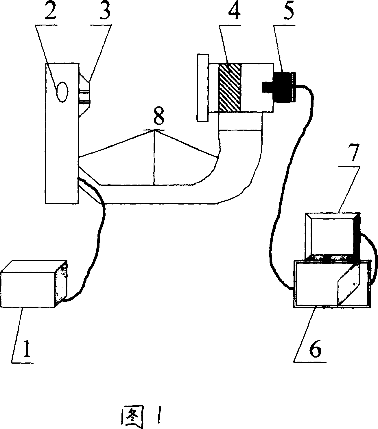
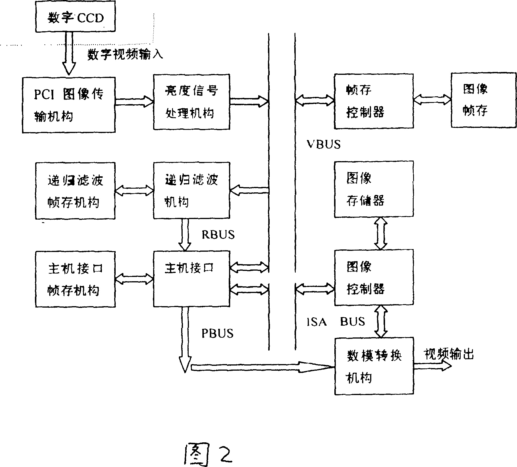
[0032] See Fig. 1 and know: the present invention mainly is made up of high-voltage generator 1, X-ray tube 2, collimator 3, image intensifier 4, digital CCD 5, computer digital imaging system 6, image display 7, mechanical support 8, wherein high-voltage generation The device 1 is connected with the X-ray tube 2 through a cable, the collimator 3 is located at the rear end of the X-ray tube 2, the X-ray tube 2 is connected with the image intensifier 4 through a mechanical bracket 8, and a digital CCD5 is installed at the rear end of the image intensifier 4 , digital CCD5 is connected with computer digital imaging system 6 by cable, and computer digital imaging system 6 is connected with image display 7 by cable, and computer digital imaging system 6 is made up of computer host computer and digital imaging device and digital imaging module device based on computer host computer, wherein The digital imaging module device controls the digital CCD5 to collect signals and inputs the...


PUM


Abstract

The digital foreign matter position finder mainly comprises a high-voltage generator, an X-ray tube, a collimating device, an image amplifier, a digital CCD, a computer digital image-forming system, an image display and a mechanical support bracket, wherein the image amplifier is provided with a digital CCD at the rear end, the digital image-forming module control the digital CCD to perform signal acquisition and input the signal to the digital image-forming device for real time treatment, and the treated signal is transmitted to the image display for displaying.

Description

technical field [0001] The invention relates to a medical device, in particular to a digital foreign object locator for detecting and locating foreign objects inside human tissues. Background technique [0002] In the past, the traditional localization methods for foreign bodies at home and abroad mainly include: 1. Anteroposterior and lateral methods: according to the anteroposterior (i.e., anteroposterior or posteroanterior) observation of metal foreign bodies located on the inside or outside, and lateral observation of metals located on the ventral or dorsal side In some cases, judge the position and depth of the foreign body, and make skin marks for surgical removal. Generally, it is necessary to use a combination of fluoroscopy and radiographs to achieve accurate positioning; For removal, first turn the patient under fluoroscopy to find the point closest to the body surface of the foreign body, then carry out local disinfection and anesthesia, th...

Claims


Application Information

Patent Timeline
Patent Type & Authority Patents(China)
IPC IPC(8): A61B6/12
Inventor 陈菁李兵仓康建毅赵辉陈志强张良潮
Owner INST OF FIELD OPERATION SURGERY NO 3 MILITARY MEDICL UNIV PLA
Why Eureka
Try Eureka