X-ray computer chromatographic camera device

A photography device, X-ray technology, applied in X-ray equipment, echo tomography, measuring devices, etc., can solve the problem of inappropriate scanning speed

Inactive Publication Date: 2003-09-10
TOSHIBA MEDICAL SYST CORP
View PDF1 Cites 14 Cited by
  • Summary
  • Abstract
  • Description
  • Claims
  • Application Information

AI Technical Summary

Problems solved by technology

Therefore, in some cases, the scan speed in the mai...

Method used


Image

  • X-ray computer chromatographic camera device
  • X-ray computer chromatographic camera device
  • X-ray computer chromatographic camera device

Examples


Embodiment Construction

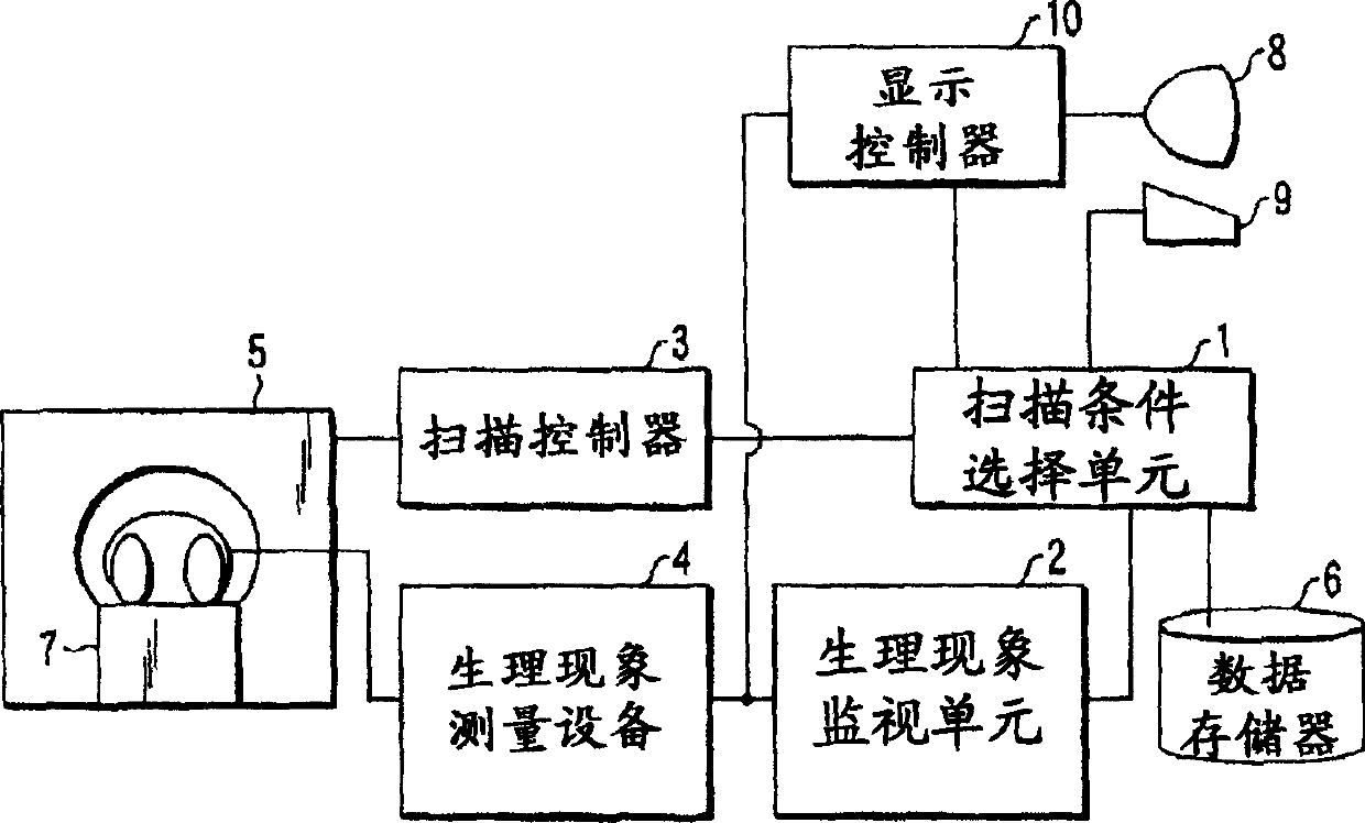
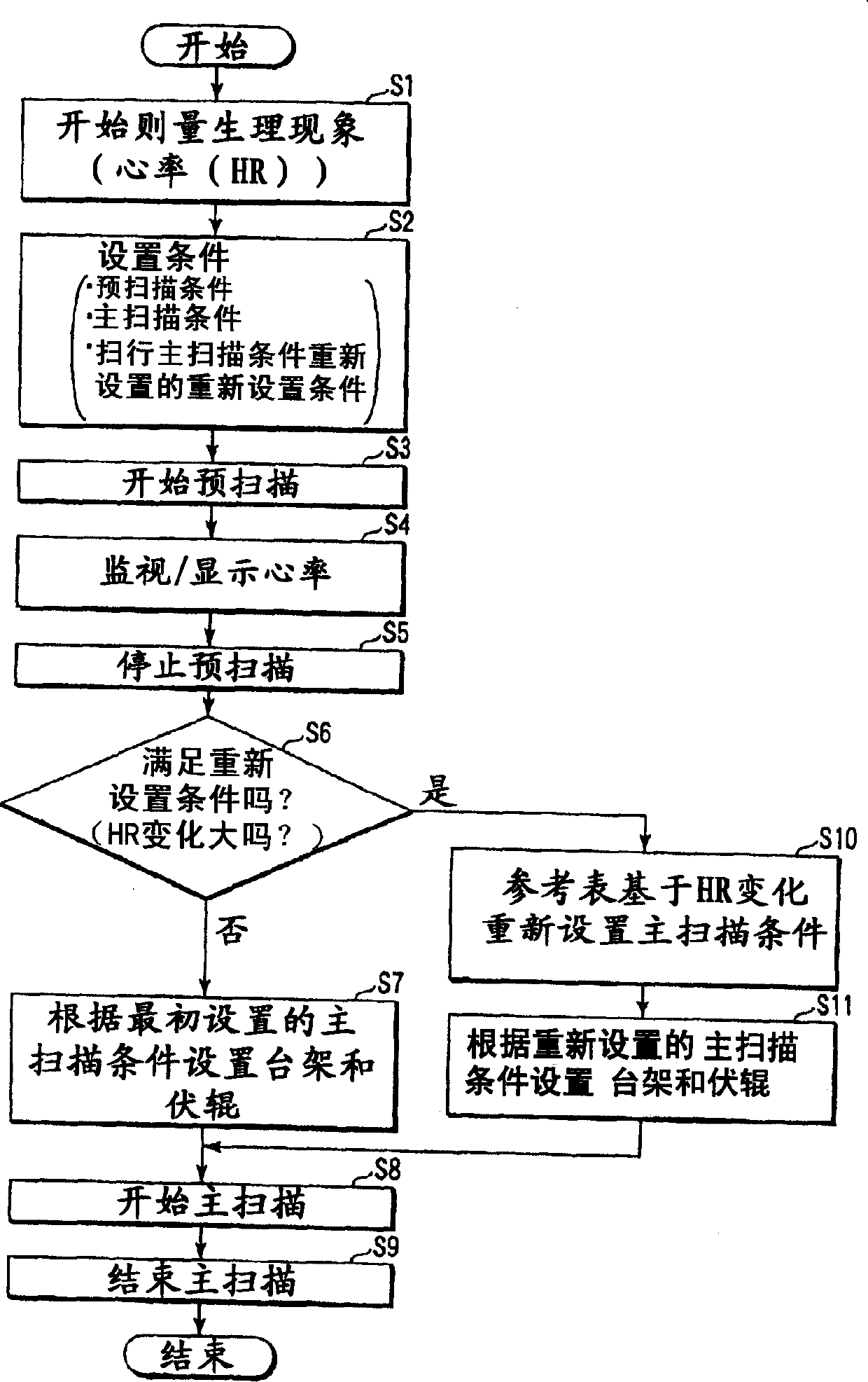
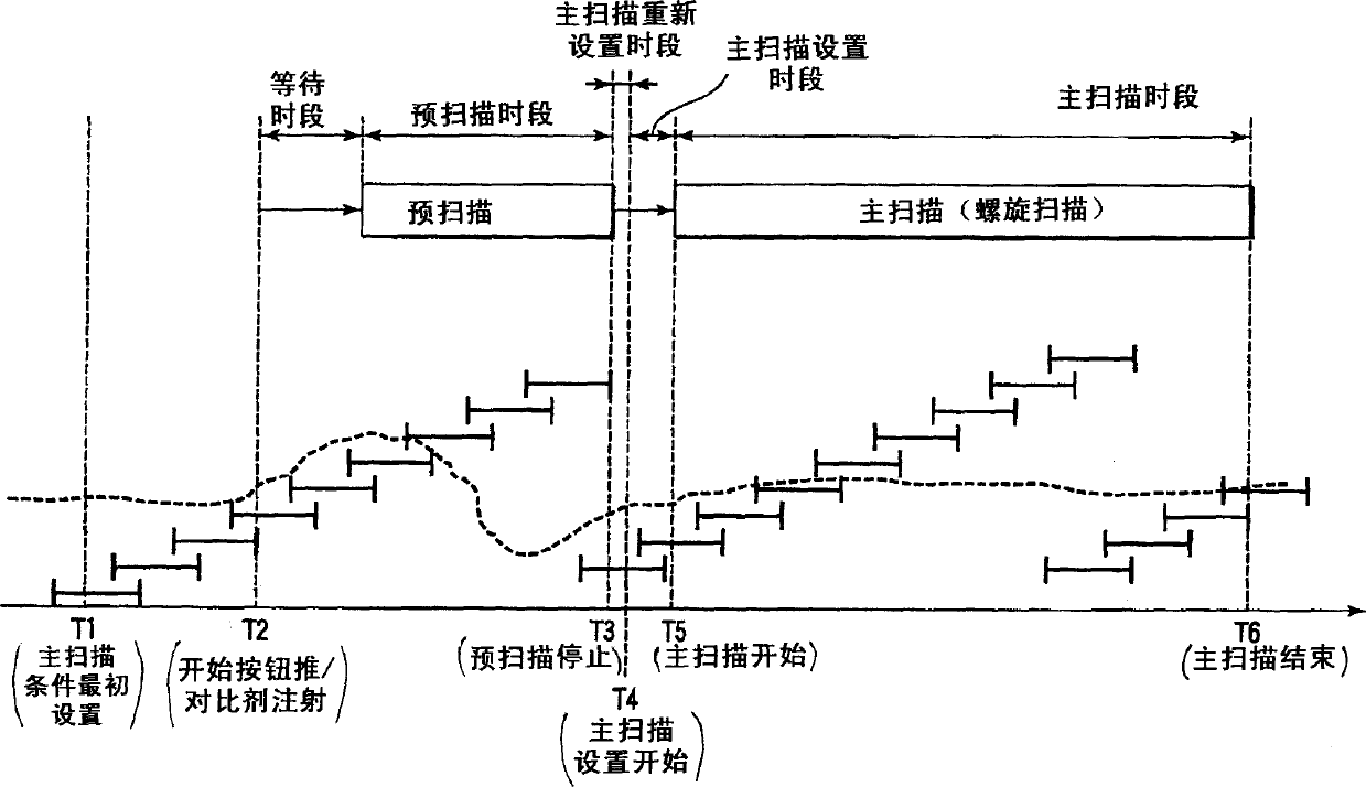
[0031] An X-ray computed tomography apparatus according to a preferred embodiment of the present invention will be described below with reference to the views of the accompanying drawings. Note that X-ray computed tomography devices include various types of devices, for example, rotary / rotary type devices in which the X-ray tube and X-ray detector rotate together around the object to be inspected; stationary / rotary Type of device in which a number of detection elements are arranged in a ring and only the x-ray tube rotates around the object to be inspected. The invention can be applied to either type. In this example, the current mainstream type - rotation / rotation type - will be taken as an example for illustration.

[0032] In addition, image reconstruction methods include a 360° method that requires 360° projection data corresponding to one rotation around an object to be inspected, and a half-scan method that requires (180°+fan angle) projection data. The present inventi...


PUM


Abstract

An X-ray computer tomography apparatus of this invention has a function of monitoring, by a pre-scan, a density of a contrast medium in ROI of an object, stopping the pre-scan on the basis of a timing at which the density reaches to a predetermined value, and starting a main-scan. A main-scan condition is initially set based on a physiological phenomenon of the object measured before the injection of the contrast medium. The necessity of resetting an initially set main scan condition is determined on the change of a physiological phenomenon measured before the end of the pre-scan. If it is determined that the main scan condition needs to be reset, the main scan condition is reset from the initially set main scan condition to a main scan condition corresponding to the physiological phenomenon measured before the end of the pre-scan.

Description

[0001] Cross References to Related Applications [0002] This application is based on and claims priority from Japanese Application Patent No. 2002-051878 filed on February 27, 2002, the entire contents of which are incorporated herein by reference. technical field [0003] The present invention relates to an X-ray computed tomography apparatus having a function of performing a pre-scan before performing a main scan. Background technique [0004] In contrast-medium imaging, it is very important to start scanning (main scanning) when a contrast medium injected into an object to be inspected flows into a relevant area. Therefore, a scan (pre-scan) is performed before the main scan. In order to suppress the X-ray dose, the pre-scan is performed by using X-rays weaker in intensity than the X-rays used in the main scan. Provides projection data acquired in pre-scan for real-time reconstruction processing. The real-time reconstructed image (tomographic image) is displayed immed...

Claims


Application Information

Patent Timeline
IPC IPC(8): A61B5/352A61B6/00
CPCY10S378/901A61B6/481A61B6/541A61B6/488A61B5/0456A61B6/032A61B6/027A61B6/504A61B6/465A61B5/352
Inventor 尾嵜真浩
Owner TOSHIBA MEDICAL SYST CORP
Why Eureka
Try Eureka