Method and apparatus for motion correction in 3-D medical image sequence

A technique of objects, sequences, applied in the field of time series and equipment for scanning moving objects, which can solve the problems of insufficient examination duration to show all phenomena, inability to eliminate organs, and inability to study vein equivalence

Inactive Publication Date: 2005-06-01
GE MEDICAL SYST GLOBAL TECH CO LLC
View PDF0 Cites 9 Cited by
  • Summary
  • Abstract
  • Description
  • Claims
  • Application Information

AI Technical Summary

Problems solved by technology

However, the duration of the examination corresponding to the period during which the patient is able to hold his breath is insufficient to show all the phenomena that occur during one blood cycle
Specifical

Method used


Image

  • Method and apparatus for motion correction in 3-D medical image sequence
  • Method and apparatus for motion correction in 3-D medical image sequence
  • Method and apparatus for motion correction in 3-D medical image sequence

Examples


Embodiment Construction

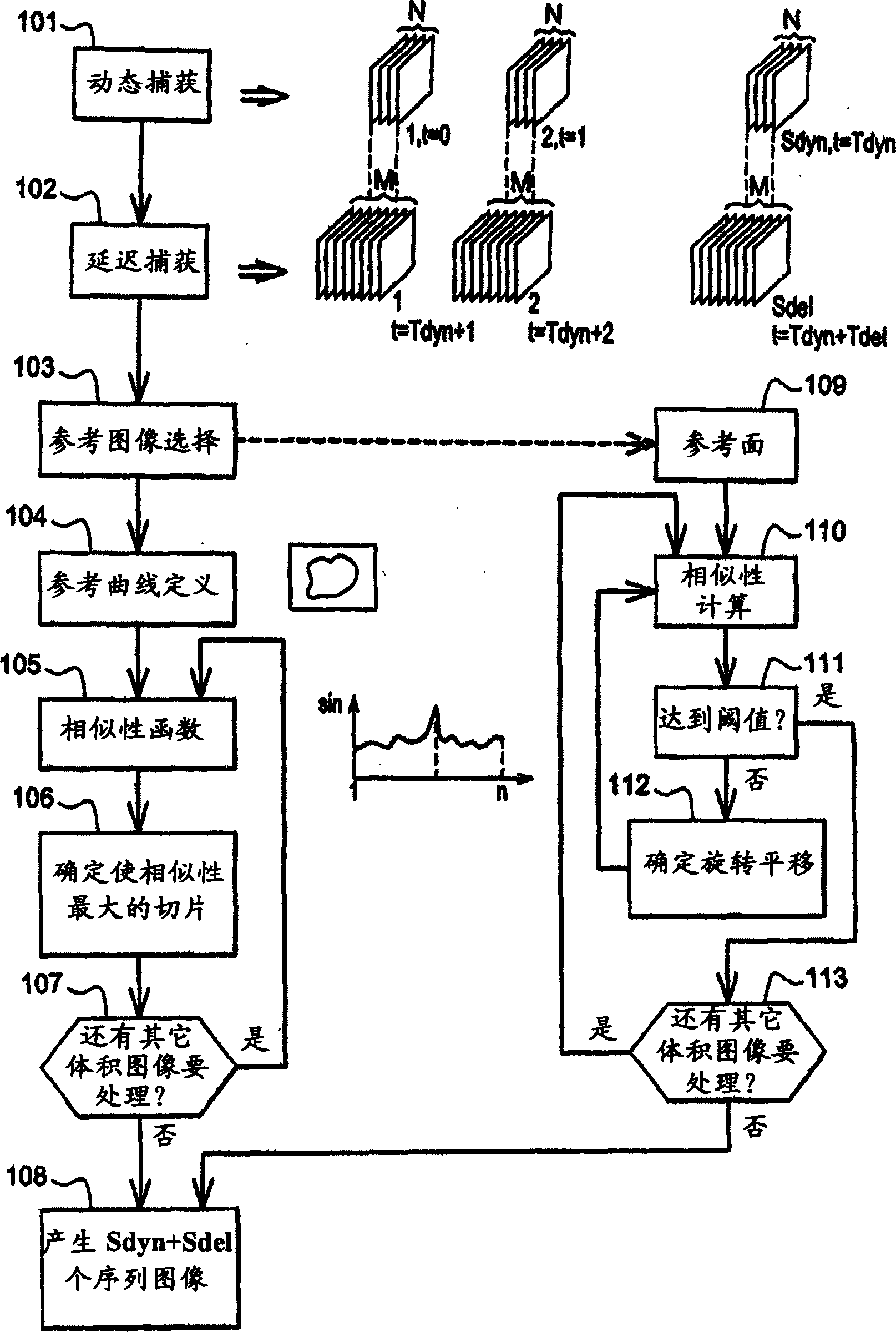
[0014] figure 1 A step 101 is shown for dynamically capturing a volumetric image from an organ in a living body. In fact, and for purposes of illustration, a living body is a human. However, the invention is equally valid for other living organisms, in fact, for all higher organisms. In this way, it is sufficient to adjust the capture parameters of the device performing the image capture.

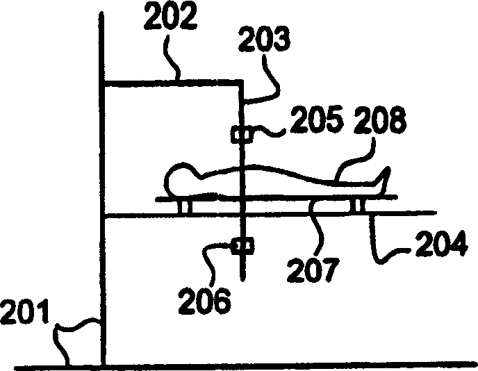
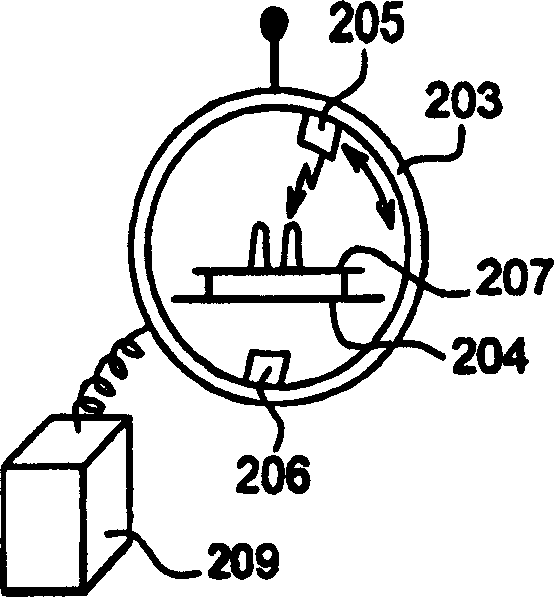
[0015] Figure 2a Schematic illustration of the volumetric image capture device. A conventional scanner is considered in the description, but in practice this device could also be a magnetic resonance device. Figure 2a A chassis 201 is shown with an arm 202 secured thereto. Underframe 201 and arm 202 provide support for capture device 203 and horizontal inspection table 204 . The capture device 203 is generally annular and the table 204 is positioned so that the patient lying on the table is in the center of the device 203 . Apparatus 203 includes an emitter 205 and a sensor 206 whi...


PUM


Abstract

In order to prolong the duration of an examination by a scanner the examination is carried out in two phases. A first dynamic phase (101) during which the patient holds his breath and the scanner explores a region Rdyn, and a second delayed phase (102) during which the patient breathes freely and the scanner explores the region Rdel. The latter comprises the region Rdyn. The images acquired during the delayed phase are registered (103-107, 109-113) with relation to at least one dynamic phase image. This registration allows an image sequence covering a long time period for region Rdyn to be obtained. This registration permits being free from the effects of the respiratory and cardiac cycles. <IMAGE>

Description

[0001] Cross References to Related Applications [0002] This application claims priority under Title 35, United States Code, Section 119(a)-(d) of French Patent Application No. 0350927, filed November 28, 2003, which is hereby incorporated by reference in its entirety middle. technical field [0003] One embodiment of the invention relates to a method and apparatus for scanning a time series of moving objects. One embodiment of the invention relates to a method and apparatus for generating a sequence of volumetric images of a region of an organ in a living body. The field of the invention is medical imaging and devices that can obtain image sequences from organs in a living body, preferably a human being. In particular, the field of the invention is tomodensitometry or conventional imaging and magnetic resonance imaging. Background technique [0004] In the prior art, several techniques including X-rays, tomodensitometry or magnetic resonance are used to perform organ ex...

Claims


Application Information

Patent Timeline
IPC IPC(8): A61B6/03A61B5/055G06T1/00G06T7/00
CPCA61B6/5288A61B6/032A61B6/504A61B6/5205A61B6/5235G06T7/30G06T2207/30004
Inventor G·卡尔蒙T·-Y·李D·德鲁蒙德K·普罗克诺夫
Owner GE MEDICAL SYST GLOBAL TECH CO LLC
Why Eureka
Try Eureka