
X-ray CT apparatus and myocardial perfusion image generating system

A CT image and X-ray technology, which is applied in the field of X-ray CT equipment and myocardial perfusion image generation systems, can solve the problems of difficulty in ensuring long imaging time of myocardial perfusion images, and limited imaging time and frequency.

Active Publication Date: 2005-12-14
TOSHIBA MEDICAL SYST CORP
View PDF2 Cites 9 Cited by
  • Summary
  • Abstract
  • Description
  • Claims
  • Application Information

AI Technical Summary

Problems solved by technology

However, there is an upper limit to the injection amount of the contrast agent with respect to the subject, and thus, the amount of time and the number of times of imaging are also limited
Therefore, this raises the problem that it is difficult to secure a long imaging time for myocardial perfusion images

Method used


Image

  • X-ray CT apparatus and myocardial perfusion image generating system
  • X-ray CT apparatus and myocardial perfusion image generating system
  • X-ray CT apparatus and myocardial perfusion image generating system

Examples


Embodiment Construction

[0029] An X-ray CT apparatus and a myocardial perfusion image generating system according to the present invention will now be described in more detail below with reference to embodiments with reference to the accompanying drawings.

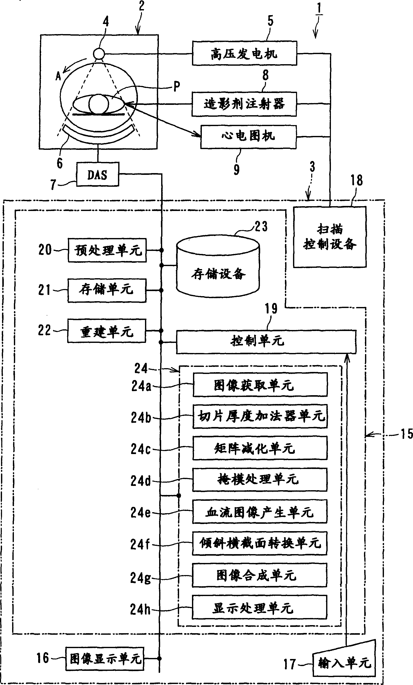
[0030] figure 1 is a configuration diagram illustrating an X-ray CT apparatus according to an embodiment of the present invention. The X-ray CT apparatus 1 includes a gantry unit 2 and computer equipment 3 . The gantry unit 2 includes an X-ray tube 4 , a high voltage generator 5 , an X-ray detector 6 , a DAS (Data Acquisition System) 7 , a contrast agent injector 8 , and an electrocardiograph 9 . The X-ray tube 4 and the X-ray detector 6 are mounted in positions facing each other sandwiching the subject P in a not-shown rotating ring that rotates continuously at high speed.

[0031] The contrast medium injector 8 controlled by a control signal from the computer device 3 has a function of continuously injecting a contrast medium into the subject...


PUM


Abstract

The present invention discloses an X-ray CT apparatus (1) for radiating X-rays to a subject (P) for scanning the subject (P) and reconstructing an image inside the subject (P) based on the obtained projection data, It includes an image generation unit and a blood flow image generation unit (24e). The image generating unit generates an image based on the projection data in a state where the concentration of the contrast agent in the myocardial portion of the subject (P) to which the contrast agent is continuously injected can be considered constant. A blood flow image generating unit (24e) generates a blood flow image by removing components of myocardial tissue from a portion of a myocardial region of an image generated by the image generating unit.

Description

[0001] Cross References to Related Applications [0002] This application is related to and incorporated by reference in Japanese Patent Application No. 2004-174364 filed on June 11, 2004 and Japanese Patent Application No. 2005-005977 filed on January 13, 2005. technical field [0003] The present invention relates to an X-ray CT (computed tomography) device and a myocardial perfusion image generation system, which obtain contrast CT images, such as coronary angiography CT images, myocardial contrast CT images, etc., by continuously injecting contrast agents, and Myocardial perfusion images were generated using the obtained contrast CT images. Background technique [0004] Regarding myocardial contrast examination using an X-ray CT apparatus, a contrast medium is injected into a subject as a patient, and a contrast CT image is collected. Subsequently, live images of the coronary arteries, endocardial luminal walls, etc. are generated from the collected contrast CT images a...

Claims


Application Information

Patent Timeline
IPC IPC(8): A61B6/03
CPCA61B6/507
Inventor 市原隆
Owner TOSHIBA MEDICAL SYST CORP
Learn More