Amelioration of macular degeneration and other ophthalmic diseases

A macular degeneration and disease technology, applied in skin diseases, sensory diseases, cardiovascular system diseases, etc.

Inactive Publication Date: 2006-12-20
COLBY PHARMA CO
View PDF30 Cites 15 Cited by
  • Summary
  • Abstract
  • Description
  • Claims
  • Application Information

AI Technical Summary

Problems solved by technology

However, apart from the highly reducing environment of the eye lens (M.Lou, 2003, supra), there have been no reports of the use of hydroxylamine compositions in the treatment of ocular diseases or disorders

Method used


Image

  • Amelioration of macular degeneration and other ophthalmic diseases
  • Amelioration of macular degeneration and other ophthalmic diseases

Examples


Embodiment 1

[0074] Example 1: Therapeutic effect of Tempol-H on photochemical retinal damage in a rabbit model

[0075] The ability of light to produce retinal damage far below the level at which it can produce thermal damage is referred to as photochemical retinal damage. It is generally believed that the mechanism of action of photochemical retinal damage is the generation of free radicals induced by light. The ability of common light sources used in clinical ophthalmology to produce photochemical retinal damage is now well appreciated. Manipulating microscopes used in ophthalmic surgery have demonstrated the ability to produce photochemical damage to the retina. This observation has been used to create a model in the rabbit eye to test the defense of various agents against actinic retinal damage. It has been determined that optically visible retinal damage is detectable after only 2.5 minutes of exposure to an operating microscope. The protocol set out below can be used to determine...

Embodiment 2

[0079] Example 2: Evaluation of Tempol-H Incorporation into Rabbit Retinal Tissue Following Intravitreal Injection

[0080] In order to evaluate the effectiveness of tempol-H in preventing AMD and other retinal disorders, it is necessary to establish that the drug has incorporated into retinal tissue. The protocol set out in this example employs scintillation techniques to determine the amount and duration of tempol-H incorporation into retinal tissue following intravitreal injection into the rabbit. Because the large eyes of rabbits provide an appropriate template for therapeutic administration, New Zealand white rabbits were characterized in detail in experiments involving intravitreal injections (Hosseini et al., 2003, Lasers Surg. Med. 32:265-270). In addition, New Zealand white rabbits have been used extensively in experiments evaluating drug uptake into retinal tissue by scintillation techniques (Ahmed et al., 1987, J. Pharm. Sci. 76:583-586).

[0081] Materials and Met...

Embodiment 3

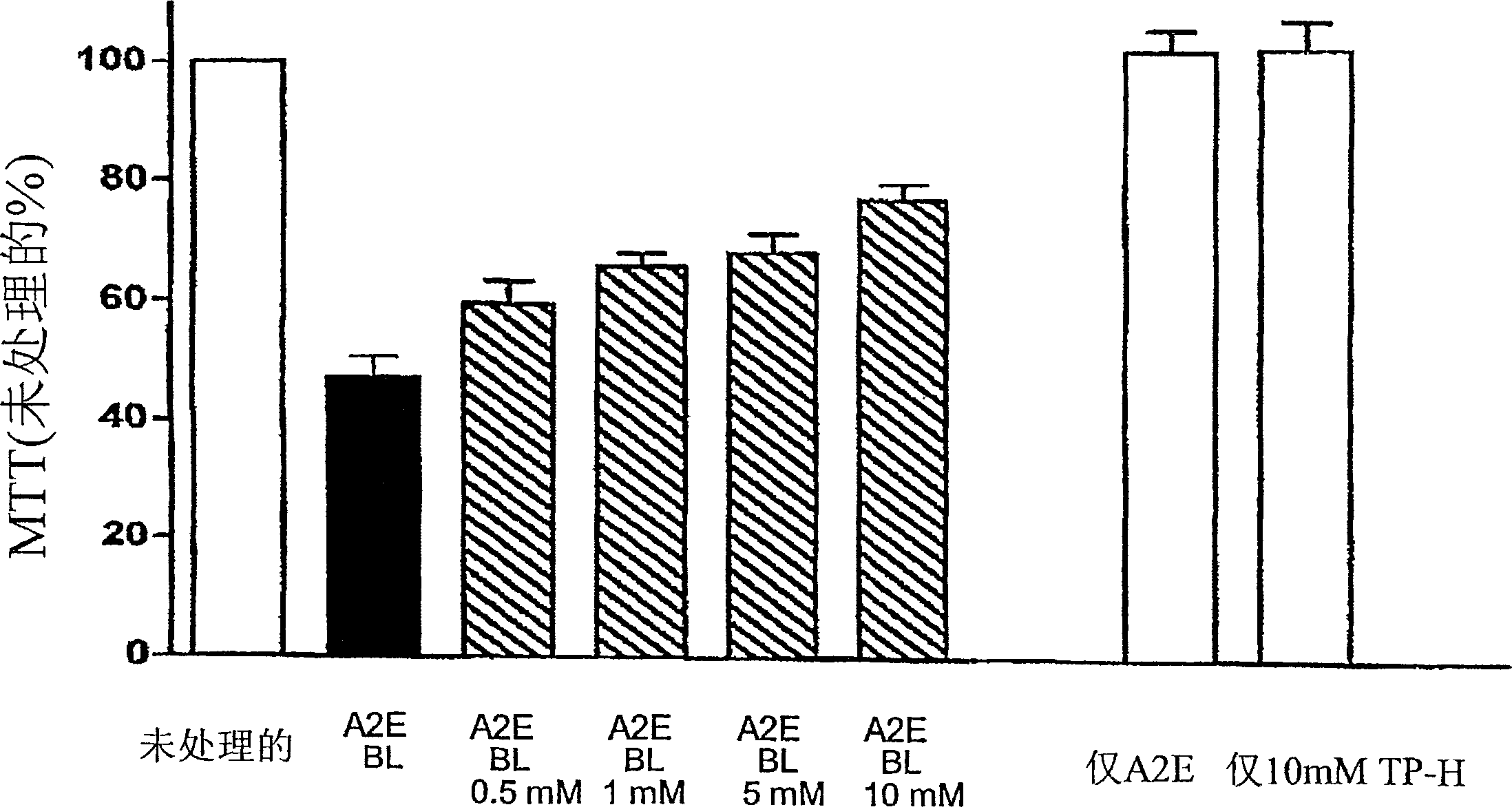
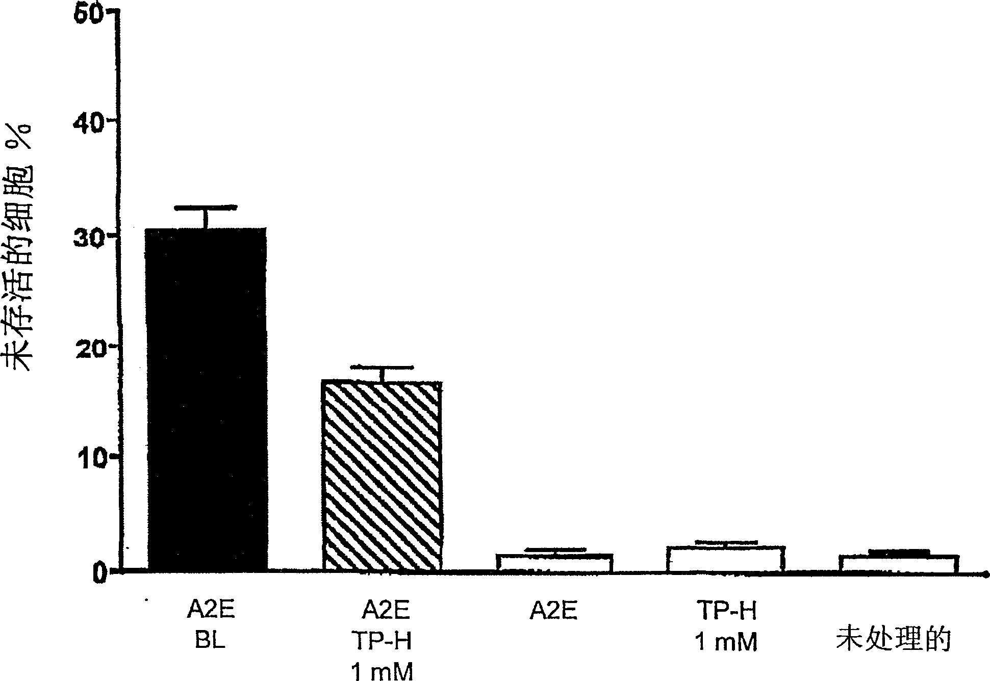
[0088] Example 3: Efficiency of Tempol-H in Protecting Photooxidative Processes in Retinal Pigment Epithelial Cells (RPE)

[0089] background:

[0090] Although the etiology of atrophic age-related macular degeneration (AMD) is unclear, it is generally accepted that AMD begins with the death of retinal pigment epithelium, degeneration of photoreceptor cells, and the consequent visual impairment lost. There is increasing evidence linking lipofuscin accumulation in the RPE to the death of these cells. For example, not only the level of lipofuscin in RPE cells below the macula is the highest, but the measurement of lipofuscin in RPE by automatic fundus fluorescence also demonstrates that at the site of previous fluorescence enhancement, the area of ​​RPE atrophy will expand accordingly. Studies on the relationship between RPE lipofuscin and RPE cell death have shown that the component of RPE lipofuscin, the bisretinoid luminophore A2E, can disrupt cell membranes and mediate blu...


PUM


Abstract

Methods for the treatment or prevention of a number of ocular diseases or disorders are disclosed. In particular, methods are disclosed for the amelioration of macular degeneration, diabetic retinophaty and other retinopathies, as well as uveitis, presbyopia, dry eye, glaucoma, blepharitis and rosacea of the eye. The methods comprise administration of a compositions comprising an ophthalmologically acceptable carrier or diluent and a hydoxylamine compound in a therapeutically sufficient amount to prevent, retard the development or reduce the symptoms of one or more of the ophthalmic conditions.

Description

[0001] Cross References to Related Applications [0002] This application claims priority to US Provisional Application 60 / 523,802, filed November 20, 2003, the entire contents of which are hereby incorporated by reference. field of invention [0003] The present invention relates to the treatment of ophthalmic diseases. More specifically, the present invention provides methods of alleviating or preventing macular degeneration and other deleterious diseases of the retina and surrounding tissues through the administration of pharmaceutical formulations comprising hydroxylamine compounds. Background of the invention [0004] Various patents and other documents are cited herein. The contents of each of these patents and documents are hereby incorporated by reference in their entirety. [0005] As a complex and sensitive organ of the body, the eye is subject to a variety of diseases and other harmful conditions that interfere with its normal function. Many of these diseases c...

Claims


Application Information

Patent Timeline
Patent Type & Authority Applications(China)
IPC IPC(8): A61K31/445A61KA61K31/13A61K31/198A61K38/05
CPCA61K31/42A61K31/13A61K31/445A61P1/00A61P17/14A61P27/00A61P27/02A61P27/04A61P27/06A61P27/08A61P27/10A61P43/00A61P9/10
Inventor 威廉·L·马蒂尔甘希亚姆·帕蒂尔
Owner COLBY PHARMA CO
Why Eureka
Try Eureka