Device, system and method for quality assessment of medical images

a medical image and quality assessment technology, applied in the field of medical image quality assessment devices, systems and methods, can solve the problems of inaccurate information, negative impact of clinical workflow, and inability to verify information, so as to improve the quality assessment of medical images and facilitate the assessment of the correctness of anatomical information

Active Publication Date: 2019-09-03
KONINKLJIJKE PHILIPS NV
View PDF25 Cites 0 Cited by
  • Summary
  • Abstract
  • Description
  • Claims
  • Application Information

AI Technical Summary

Benefits of technology

[0006]It is an object of the present invention to provide a device, system and method for improved quality assessment of medical images, in particular for easy and fast assessment of the correctness of anatomical information depicted in an acquired medical image.
[0030]In an embodiment said analysis unit is further configured to compare the obtained medical image with the reference image to generate quality information representing a level of similarity. This provides a simple way of obtaining quality information using only image comparison techniques.
[0032]The analysis unit may further be configured to use an imaging guideline including attributes, in particular DICOM attributes, and / or imaging planning guidelines in the analysis to generate said quality information representing compliance and / or the level of compliance of the obtained medical image with the attributes and / or imaging planning guidelines. This further improves the reliability of the assessment.
[0033]In another embodiment the device further comprises an image processing unit configured to detect target anatomy and / or anatomical landmarks in the obtained medical image, wherein said analysis unit is further configured to compare the detected target anatomy and / or anatomical landmarks in the obtained medical image with target anatomy and / or anatomical landmarks in the reference image to generate quality information representing a level of similarity. The use of target anatomy and / or anatomical landmarks allows a precise analysis and assessment.

Problems solved by technology

Another (even more fundamental) issue is the correctness of anatomical information.
While the Digital Imaging and Communications in Medicine (DICOM) standard provides multiple options to indicate the scanned anatomy (e.g. Body Part Examined value or Anatomic Region Sequence code), this information is not reliable.
Errors in the anatomical information could have a negative impact on the clinical workflow, as e.g. relevant prior scans are more difficult to identify.
Furthermore, it makes a statistical evaluation of the performed scans in a department impossible.

Method used


Image

  • Device, system and method for quality assessment of medical images
  • Device, system and method for quality assessment of medical images

Examples


Embodiment Construction

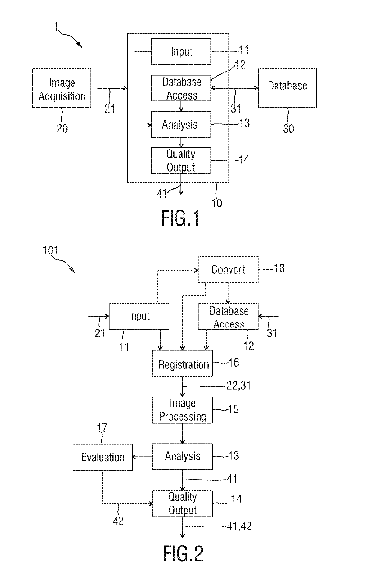
[0043]FIG. 1 shows a schematic diagram of an embodiment of a system 1 and device 10 for quality assessment of medical images according to the present invention. In addition to the device 10 the system comprises an image acquisition device 20 configured to acquire medical images of a subject according to an imaging guideline. The image acquisition device 20 may generally be of any kind, which allows acquisition of a medical image, such as an x-ray device, a CT device, an ultrasound device, an MR device, a PET device, a SPECT device, etc.

[0044]Based on the image data, which may e.g. a single image, a set of images, a two- or three-dimensional image data set, etc., the device 10 assesses their quality acquired with said image acquisition device 20 according to an imaging guideline. Such an imaging guideline may be prescribed by a manual of the imaging guideline, or by a guideline, by a standard, by a physician, etc. and defines which kind if image data shall be acquired. For instance, ...


PUM


Abstract

The present invention relates to a device, system and method for quality assessment of medical images. The device comprises an image input configured to obtain a medical image acquired according to an imaging guideline, a database access unit configured to access a database storing reference images for a plurality of imaging guidelines and for obtaining a reference image based on the imaging guideline used for acquisition of the obtained medical image, an analysis unit configured to analyze the obtained medical image in view of the obtained reference image and / or the used imaging guideline to generate quality information representing the quality of the obtained medical image, and a quality output configured to output the generated quality information.

Description

FIELD OF THE INVENTION[0001]The present invention relates to a device, system and method for quality assessment of medical images.BACKGROUND OF THE INVENTION[0002]Worldwide there is an increased pressure on imaging departments to measure and improve the quality of care. In the United States, this is driven by regulatory agencies, the Centers for Medicare and Medicaid Services (CMS), payers as well as professional societies.[0003]Common key performance indicators (KPIs) are based on quantities such as equipment utilization, idle time or patient throughput. By now, even first benchmarks for quality improvement became available which allow for a comparison between different sites. Most of the KPIs are derived from device logs or clinical information systems (i.e. non-image data).[0004]Next to non-image data, the images themselves provide a broad range of options to define meaningful quality metrics. One example is the adherence to guidelines. Imaging departments typically define scan p...

Claims


Application Information

Patent Timeline
Patent Type & Authority Patents(United States)
IPC IPC(8): G06K9/00G06T7/00G06T1/00
CPCG06T7/0014G06T7/0012G16H30/20G06T1/0007G06T2207/10004G06T2207/30168G16H70/20G16H30/40
Inventor SAALBACH, AXELBYSTROV, DANIELBUELOW, THOMASLU, KONGKUO
Owner KONINKLJIJKE PHILIPS NV
Why Eureka
Try Eureka