

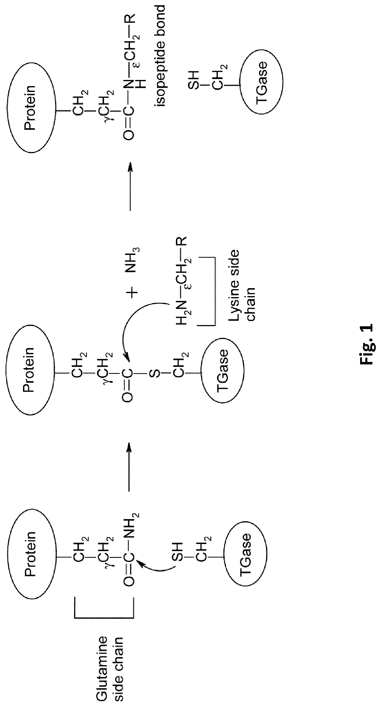
Monoclonal antibody specific for gamma-glutamyl-l-epsilon-lysine for the monitoring of apoptosis
a technology of gamma-glutamyl-l-epsilon-lysine and monoclonal antibody, which is applied in the field of monoclonal antibody specific for gamma-glutamyl-l-epsilon-lysine for the monitoring of apoptosis, can solve the problems of polyamine crosslinks being likely to interfere with detection, enzymes not activated by normal, and not readily availabl
- Summary
- Abstract
- Description
- Claims
- Application Information
AI Technical Summary
Benefits of technology
Problems solved by technology
Method used
Image


Examples
example
Example 1: Development of a Monoclonal Antibody Specific to Gamma-Glutamyl-L-Epsilon-Lysine (GGEL)
[0231]Materials & Methods
[0232]Development of mAb, Preparation of Immunogen and Immunization Regime.
[0233]Mice were immunized with gamma-glutamyl-L-epsilon-Lysine (GGEL) coupled on Keyhole limpet hemocyanin (KLH) via glutaraldehyde and then dialysed against Phosphate Buffer Saline 1×(PBS).
[0234]25 μg of antigenic solution was dissolve in 100 μL of PBS. Six-weeks-old BALB / c and SJL female white mice were given four intranodal injection of immunogen at 1 week intervals and a single intraperitoneal injection three days before fusion.
[0235]Production and Screening of Hybridomas.
[0236]Hybridoma cells were produced by the method described elsewhere Galfre and Milstein (1981). The supernatant were screened by enzyme-linked immunosorbent assay (ELISA) against GGEL coupled to bovine Serum Albumine (BSA) immobilized to the wells of Maxisorp microtiter plates. Wells containing immobilized antigens...
example 2
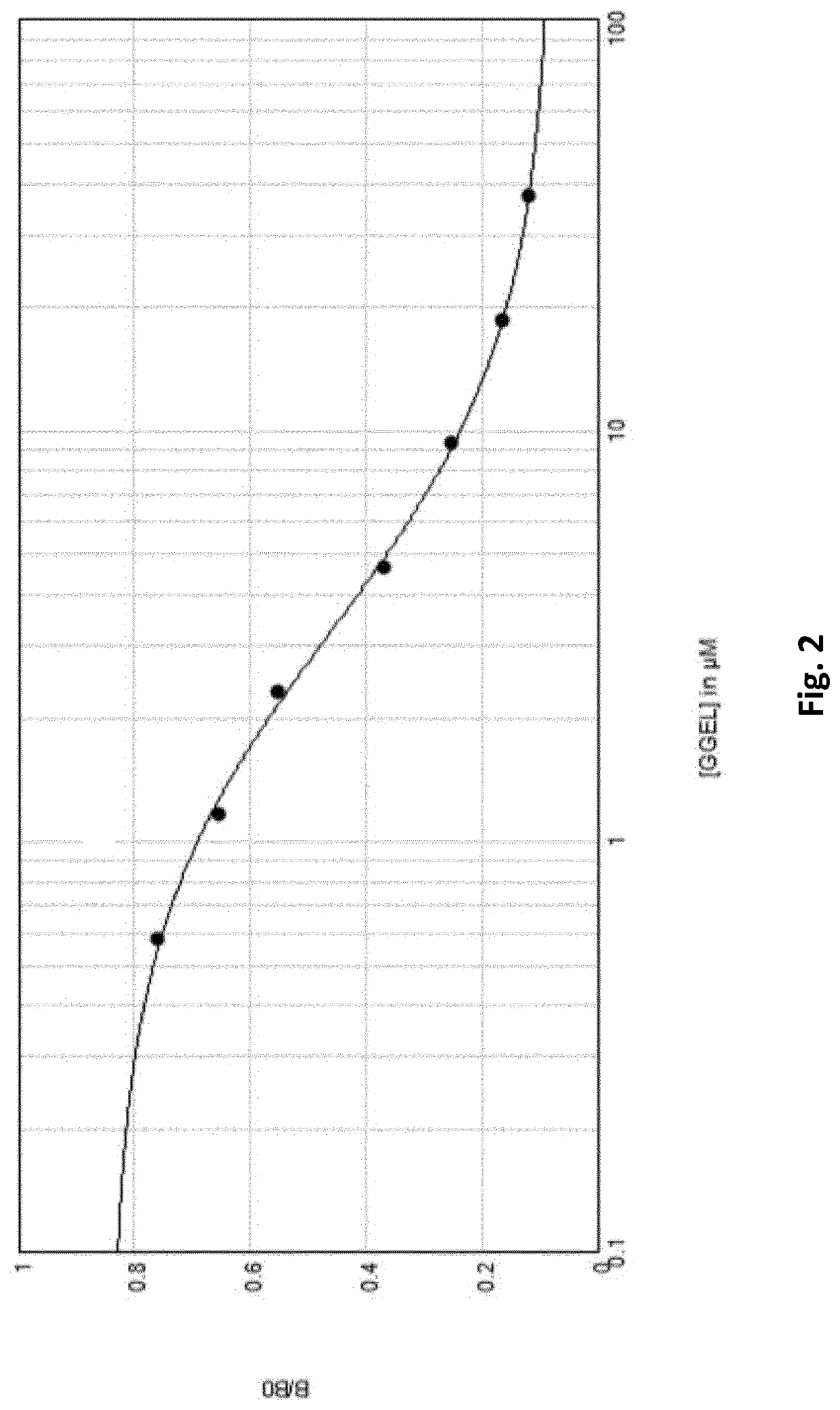
ation of Gamma-Glutamyl-L-Epsilon-Lysine (GGEL) with the 1G1h1 Monoclonal Antibody
[0268]Materials & Methods
[0269]GGEL Quantification by Competitive ELISA. Competitive ELISA.
[0270]BSA-GGEL was adsorbed on microtiter plate at a concentration of 10 μg / mL in a 50 mM bicarbonate solution pH 9.50. Plates were incubated overnight at laboratory temperature. Saturation was performed with a phosphate buffer 0.1 M supplemented of BSA 0.5% and sucrose 5% (FIG. 3, step 1). Samples diluted were added in presence of antibody solution for 1 hour at 37° C., BZGO with a precise number of GGEL “coated” on were diluted two by two and used as standard (FIG. 3, step 2 and 3). After three washes with PBST, secondary antibody diluted 1 in 2000 PBST was incubated 30 minutes at 37° C. Revelation were performed using TMB for 5 minutes and reaction were stopped using H2SO4, 2N (FIG. 3, step 4). Absorbance values were determined at 450 nm with Spectramax i3® automated microplate reader (Molecular Devices, Sunny...
example 3
Detection of Free GGEL Released by Apoptotic Cells
[0277]Materials & Methods
[0278]Cell Lines.
[0279]Cell line used for apoptosis induction was Human promyelocytic leukemia cells, HL-60. HL-60 were cultured in RPMI media supplemented with 10% Fetal Calf Serum (FCS), 1% Streptomycin / Penicillin and 200 mM L-Glutamine.
[0280]Induction of apoptosis.
[0281]Cells were plated at 1×10E6 cells / mL in cell culture medium described before. Induction of apoptosis were done using staurosporine at a concentration of 1 μmol / L during 8 hours. Staurosporine is an alkaloid isolated from the culture broth of Streptomyces staurosporesa. It is a potent, cell permeable protein kinase C inhibitor and other kinases such as PKA, PKG, CAMKII and Myosin light chain kinase (MLCK). At 0.2-1 μM, staurosporine induces cell apoptosis. After treatment, cells were then centrifuged at 800 g, 10 min and supernatant were used for GGEL quantification.
[0282]GGEL Quantification
[0283]GGEL quantification by competitive ELISA. Com...
PUM
| Property | Measurement | Unit |
|---|---|---|
| pH | aaaaa | aaaaa |
| concentration | aaaaa | aaaaa |
| humidity | aaaaa | aaaaa |
Abstract
Description
Claims
Application Information