
Quantification of absolute blood flow in tissue using fluorescence-mediated photoplethysmography

a technology of fluorescence-mediated photoplethysmography and absolute blood flow, which is applied in the field of optical assessment of blood flow in tissue using photoplethysmography, can solve the problems of ppg not being used to provide measurements in standardized units when assessing blood flow, and the quantitative assessment of tissue perfusion remains elusiv

Active Publication Date: 2020-04-28
STRYKER EUROPEAN OPERATIONS LIMITED
View PDF565 Cites 4 Cited by
  • Summary
  • Abstract
  • Description
  • Claims
  • Application Information

AI Technical Summary

Problems solved by technology

To date, quantitative assessment of tissue perfusion has remained elusive.
Despite the relatively wide application of PPG technology to such medical devices, PPG has not been utilized to provide measurements in standardized units when assessing blood flow.

Method used


Image

  • Quantification of absolute blood flow in tissue using fluorescence-mediated photoplethysmography
  • Quantification of absolute blood flow in tissue using fluorescence-mediated photoplethysmography
  • Quantification of absolute blood flow in tissue using fluorescence-mediated photoplethysmography

Examples


Embodiment Construction

[0028]Reference will now be made in detail to implementations and embodiments of various aspects and variations of the invention, examples of which are illustrated in the accompanying drawings.

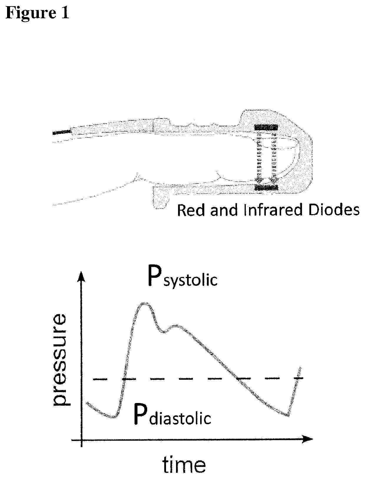
[0029]Conventional photoplethysmography (PPG) can estimate changes in tissue blood volume by detecting changes in the amount of red or near-infrared light transmitted through the tissue. As the blood volume within tissue expands and contracts during a cardiovascular pressure pulse corresponding to the heartbeat of the subject, the amount of light absorbed by the blood volume increases and decreases, respectively. As shown in FIG. 1, for example, the aggregate blood volume in the fingertip blood vessels is smallest during cardiovascular pressure pulse diastole and the volume is greatest during systole. Although it may be used for measuring pulse rate and blood oxygenation, this application of PPG technology is not configured to provide volumetric flow measurements in standardized units.

[0030]To...


PUM


Abstract

A method, an apparatus, and a kit including the apparatus and a fluorescence agent are provided for measuring a time-varying change in an amount of blood in a tissue volume, and include exciting a fluorescence agent in the blood, acquiring a time-varying light intensity signal during a pulsatile flow of the blood through the tissue volume, the pulsatile flow having a systolic and a diastolic phase resembling a conventional photoplethysmogram, and processing the acquired signal by applying a modified Beer-Lambert law to obtain a measurement of the time-varying change in the amount of blood in the tissue volume. The instantaneous molar concentration of the fluorescence agent is determined by utilizing a concentration-mediated change in a fluorescence emission spectrum of the fluorescence agent. There is further provided a fluorescence agent for use in the method.

Description

REFERENCE TO RELATED APPLICATIONS[0001]This application is a national stage application under 35 USC 371 of International Application No. PCT / IB2014 / 065189, filed Oct. 9, 2014, the entire contents of which are incorporated herein by reference.TECHNICAL FIELD OF THE INVENTION[0002]The present invention relates generally to the field of optical assessment of blood flow in tissue using photoplethysmography (PPG), and in particular to the quantitative assessment of blood flow in tissue, including microvascular blood flow in tissue.BACKGROUND OF THE INVENTION[0003]Perfusion refers to the flow of blood into and out of the tissue capillary bed. Quantification of tissue perfusion is of interest to clinicians across many surgical and non-surgical specialties. Although simple binary assessment (flow versus no-flow) may be adequate for some clinical applications, quantification of perfusion in standard measures is desirable in many other clinical applications. To date, quantitative assessment ...

Claims


Application Information

Patent Timeline
Patent Type & Authority Patents(United States)
IPC IPC(8): A61B5/026A61B5/00A61B5/0275A61B5/0295
CPCA61B5/0071A61B5/0275A61B5/0295A61B5/0261A61B5/6826A61B5/00
Inventor FLOWER, ROBERT W.STEAD, ROBERT ANTHONYBAILEY, ARTHUR E.
Owner STRYKER EUROPEAN OPERATIONS LIMITED
Learn More