Method and apparatus for identifying an organ structure of an examined object in magnetic resonance image data

a technology of magnetic resonance image data and organ structure, which is applied in the field of method for identifying the organ structure of an examined object in magnetic resonance image data, can solve the problem that the magnetic resonance measurement data are not typically directly available to a diagnostician, and achieve the goal of improving facilitating the contouring of the organ structure, and facilitating the contouring

Active Publication Date: 2020-07-07
SIEMENS HEALTHCARE GMBH
View PDF12 Cites 17 Cited by
  • Summary
  • Abstract
  • Description
  • Claims
  • Application Information

AI Technical Summary

Benefits of technology

[0015]The inventive procedure is based on coordinating the sampling scheme of k-space with the subsequent identification of the organ structure. In this way, acquisition of the magnetic resonance measurement data can be optimized such that the organ structure can be identified particularly simply or precisely in the magnetic resonance image data reconstructed from the magnetic resonance measurement data. The magnetic resonance measurement data are preferably entered in k-space in such a manner that the organ structure can be segmented and / or contoured particularly simply or precisely in the magnetic resonance image data reconstructed from the magnetic resonance measurement data. The sampling scheme of k-space can be coordinated specifically to the organ structure to be identified. The magnetic resonance sequence employed for the purpose of acquiring the magnetic resonance measurement data can specify a sampling scheme that results in particularly suitable magnetic resonance measurement data for the subsequent identification of the organ structure. Alternatively or additionally, the sequence can specify to be used as settings for the execution of the magnetic resonance sequence by the scanner, so that the sampling scheme generates particularly suitable magnetic resonance measurement data for the subsequent identification of the organ structure based on the parameters.
[0049]In this embodiment of the inventive procedure, it is possible to ensure an adequate quality of the magnetic resonance measurement data acquired by the sampling scheme for employment in the course of a treatment of the patient. A visual quality assurance is achieved in this regard with respect to the further magnetic resonance image data, which may be acquired with the use of a conventional sampling scheme. The quality assurance can ensure, for example, that the spatial geometry of the organ structure was not distorted in any relevant manner by the sampling scheme used. Quality assurance of the organ structure segmented and / or contoured on the basis of the magnetic resonance image data is also conceivable with respect to an atlas containing a typical appearance of the organ structure for a population.

Problems solved by technology

The magnetic resonance measurement data are therefore not typically directly available to a diagnostician for the purpose of making a medical diagnosis.

Method used


Image

  • Method and apparatus for identifying an organ structure of an examined object in magnetic resonance image data
  • Method and apparatus for identifying an organ structure of an examined object in magnetic resonance image data
  • Method and apparatus for identifying an organ structure of an examined object in magnetic resonance image data

Examples


first embodiment

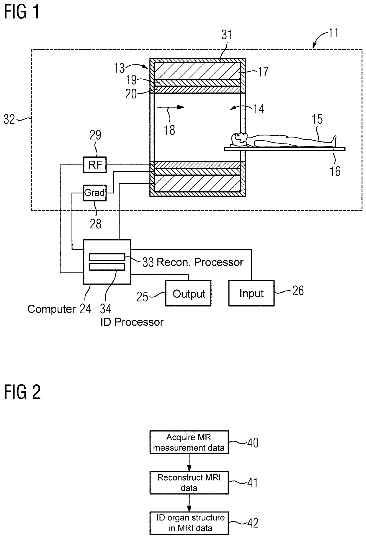
[0067]FIG. 2 shows a flowchart of the inventive method.

[0068]In a first method step 40 the scanner 13 of the magnetic resonance apparatus 11 is operated to, acquire magnetic resonance measurement data for an organ structure of the examined object using a magnetic resonance sequence that specifies a sampling scheme of k-space.

[0069]In a further method step 41 the reconstruction processor 33 of the computer 24 reconstructs the magnetic resonance image data from the magnetic resonance measurement data.

[0070]In a further method step 42 the identification processor 34 of the computer 24 identifies of the organ structure in the magnetic resonance image data.

[0071]The sampling scheme of k-space supports the subsequent identification of the organ structure in the magnetic resonance image data reconstructed from the magnetic resonance measurement data.

second embodiment

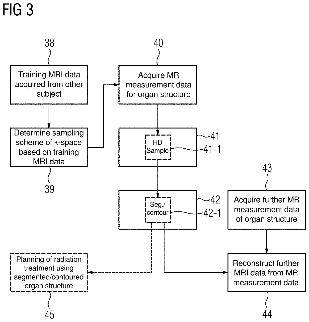
[0072]FIG. 3 shows a flowchart of an inventive method.

[0073]The following description is substantially limited to the differences from the exemplary embodiment in FIG. 2, with reference being made to the description of the exemplary embodiment in FIG. 2 in relation to identical method steps. Method steps that are substantially the same are basically numbered with the same reference numerals.

[0074]The embodiment of the inventive method shown in FIG. 3 substantially comprises the method steps 40, 41, 42 of the first embodiment of the inventive method according to FIG. 2. In addition, the embodiment of the inventive method shown in FIG. 3 comprises additional method steps and / or substeps. An alternative procedure to that in FIG. 3, which has only some of the additional method steps and / or substeps shown in FIG. 3, is also conceivable. Of course an alternative procedure to that in FIG. 3 can also have additional method steps and / or substeps.

[0075]There are different possibilities how th...


PUM


Abstract

In a method and apparatus for identifying an organ structure of an examined object in magnetic resonance image data, magnetic resonance measurement data for the organ structure of the examined object are acquired by operation of a magnetic resonance scanner using a magnetic resonance sequence that specifies a sampling scheme of k-space. Magnetic resonance image data are reconstructed from the magnetic resonance measurement data. The organ structure is identified in the magnetic resonance image data. The sampling scheme of k-space is selected so as to support the subsequent identification of the organ structure in the magnetic resonance image data reconstructed from the magnetic resonance measurement data.

Description

BACKGROUND OF THE INVENTIONField of the Invention[0001]The invention relates to a method for identifying an organ structure of an examined object in magnetic resonance image data, as well as a magnetic resonance apparatus, and a data storage medium for implementing such a method.Description of the Prior Art[0002]In a magnetic resonance apparatus, also referred to as a magnetic resonance tomography system, the body to be examined, of an examined object, for example of a patient, a healthy volunteer, an animal or a phantom, is usually exposed with the use of a basic field magnet to a relatively high basic magnet field, of 1.5 or 3 or 7 tesla for example. Additionally, gradient switching is activated with the use of gradient coils. Via a radio-frequency antenna, radio-frequency pulses, for example excitation pulses, are omitted that result in the nuclear spins of certain atoms being excited in a resonant manner by these radio-frequency pulses so as to be tilted over by a defined flip a...

Claims


Application Information

Patent Timeline
Patent Type & Authority Patents(United States)
IPC IPC(8): A61B5/055G01R33/48G01R33/56G01R33/54A61B5/00A61N5/10G01R33/58
CPCA61B5/055G01R33/5608G01R33/4818G01R33/543A61B2576/00A61B5/7257A61N5/1039A61N2005/1055G01R33/583G01R33/4824G16H30/40
Inventor AMIES, CHRISTOPHER JUDEHENGERER, ARNESCHNEIDER, RAINER
Owner SIEMENS HEALTHCARE GMBH
Why Eureka
Try Eureka