Innate metabolic imaging of cellular systems

a metabolic imaging and cellular system technology, applied in the field of image processing techniques, can solve the problems of current ability to non-invasively, critical gap in the understanding of immune responses, and bias in the measurement of tam infiltration

Active Publication Date: 2022-10-11
MEMORIAL SLOAN KETTERING CANCER CENT
View PDF16 Cites 0 Cited by
  • Summary
  • Abstract
  • Description
  • Claims
  • Application Information

AI Technical Summary

Benefits of technology

[0008]In various embodiments, the approach described herein is referred to as FeMRI. The FeMRI technique exploits the high affinity of macrophages for iron(III) to quantitatively monitor TAM infiltration in vivo without contrast agents. In certain embodiments, the FeMRI technique creates an iron(III) map that covers a discrete quantifiable concentration range at a given magnetic field. The sizes of the concentration ranges are magnetic field dependent, and the detected iron concentration ranges can be divided into an arbitrary number of sub-ranges. Corresponding pixels in these ranges are stratified from the FeMRT maps, and the location and size of pixel clusters having corresponding iron concentrations is measured. The technique has been validated with quantitative histology, and used to measure infiltration of TAMs in orthotopic models of prostate cancer during iron(III) chelation therapy, as described herein. The TAM number was found to be higher in larger tumors, but chelation was able to reduce TAMs and achieve impressive reductions in tumor growth. These results demonstrate the ability to monitor macrophages and their immune response to cancer therapy with the non-perturbing MM approach described herein, and modulate tumor growth and TAM infiltration by targeting their innate metabolism of iron.

Problems solved by technology

However, iron contrast agents can hyperpolarize the iron metabolism of macrophages promoting a pro-tumor phenotype, therefore biasing the measurement of TAM infiltration.
Thus, current ability to non-invasively assess TAM number, and monitor TAM response over the full course of therapeutic regimens using MRI is limited, leaving a critical gap in the understanding of immune response to cancer therapy and how to enhance it further.
This is because label-based MRI approaches are hampered by the contrast agent itself due to various factors, such as variable cell uptake and limited lifetime of the cellular label (<1 week).

Method used


Image

  • Innate metabolic imaging of cellular systems
  • Innate metabolic imaging of cellular systems
  • Innate metabolic imaging of cellular systems

Examples


experiment b

AM Infiltration during Prostate Cancer Therapy

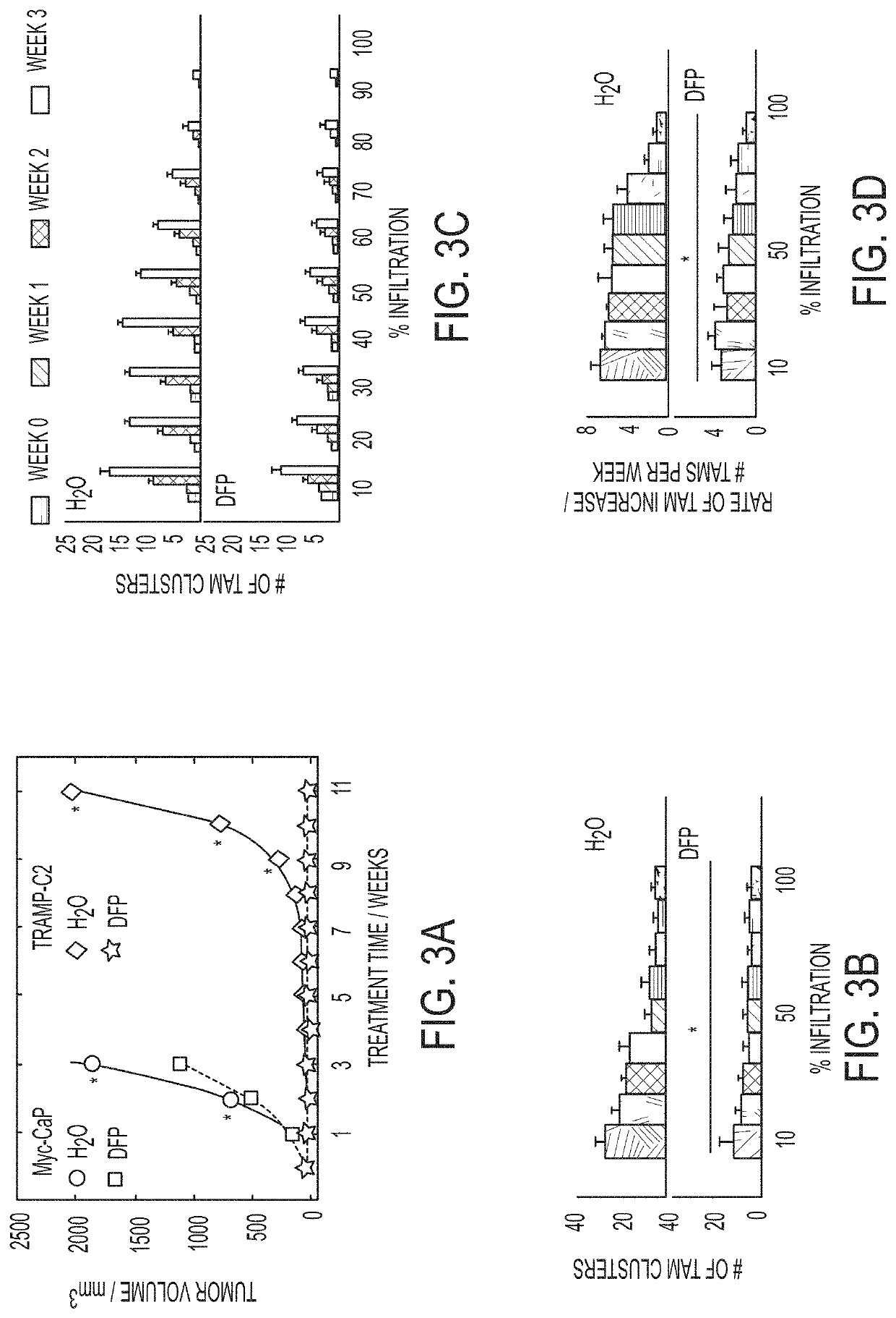
[0092]Preliminary data presented below indicates that FeMRI and histology can be used in combination with iron chelation therapy to monitor reductions in TAM infiltration that coincide with significant inhibition of prostate tumor growth in immune competent orthotopic models. This therapeutic effect resembles macrophage depletion caused by immune therapies such as CSF1R inhibition that also have succeeded in reducing tumor growth, and sensitize tumors to radiation. Thus, FeMRT and histology can be used to monitor TAM infiltration during (1) radiation, (2) CSF1R targeted immune therapy, and (3) iron chelation. Without wishing to be bound to any particular theory, it is believed that depletion of TAMs by immune therapy and chelation will inhibit tumor growth and sensitize tumors to radiation. The biomarker potential of TAM infiltration can lead to improved predictions of therapeutic response using a highly translatable combination of FeMRT...


PUM


Abstract

Described herein are systems and methods for image-based (e.g., MRI-based) spatial and temporal mapping of macrophages and other cell types, without the need for image contrast agents. These systems and methods are particularly useful for imaging macrophages because they naturally store metabolites, such as iron. Alternatively, the systems and methods described herein can be used where contrast agents are administered, rather than looking only at endogenous metabolite deposits.

Description

CROSS-REFERENCE TO RELATED APPLICATION[0001]This application claims the benefit of U.S. Application Ser. No. 62 / 320,347 filed on Apr. 8, 2016, the disclosure of which is hereby incorporated by reference in its entirety.GOVERNMENT SUPPORT[0002]This invention was made with government support under grant number W81XWH-13-1-0386 awarded by Department of Defense (DOD). The government has certain rights in the invention.FIELD OF THE INVENTION[0003]This invention relates generally to image processing techniques. In particular embodiments, the invention relates to processing of an MM image for mapping of cell types based on their endogenous iron concentrations.BACKGROUND[0004]Stromal infiltration of macrophages is implicated in the progression of many types of cancer, including prostate cancer (PCa). Depletion of tumor-associated macrophages (TAMs) inhibits tumor growth and enhances radiation therapy response. An important factor determining macrophage infiltration is tissue iron load becau...

Claims


Application Information

Patent Timeline
Patent Type & Authority Patents(United States)
IPC IPC(8): A61B5/00G06T7/11G06T7/00G16H30/20A61B5/055A61B5/145A61B6/03A61B8/08G06T15/08
CPCA61B5/4848A61B5/0035A61B5/0036A61B5/0071A61B5/055A61B5/14546A61B5/4866A61B5/7264A61B6/032A61B6/037A61B8/085G06T7/0012G06T7/0016G06T7/11G16H30/20G06T15/08G06T2207/10064G06T2207/10081G06T2207/10088G06T2207/10104G06T2207/10108G06T2207/10121G06T2207/10132G06T2207/30024G06T2207/30096G06T2210/41
Inventor LEFTIN, AVIGDORKOUTCHER, JASON
Owner MEMORIAL SLOAN KETTERING CANCER CENT
Why Eureka
Try Eureka