
Implantable devices and methods to treat benign prostate hyperplasia (BPH) and associated lower urinary tract symptoms (LUTS)

a technology of benign prostate and implantable devices, which is applied in the field of implantable devices and methods to treat benign prostate hyperplasia and associated lower urinary tract symptoms (luts), can solve the problems of many of the problems experienced, the thicker and weaker bladder wall of bph patients, and the inability of bph patients to completely empty the bladder

Active Publication Date: 2022-11-15
PRODEON MEDICAL CORP
View PDF37 Cites 0 Cited by
  • Summary
  • Abstract
  • Description
  • Claims
  • Application Information

AI Technical Summary

Benefits of technology

[0024]The implant performance criteria include expansion with sufficient force to engage and or retract tissue at the lobes of the prostate, and depending on the specific physiology of a patient, engage and displace the lobes of the prostate, thereby increasing the diameter of the urethra for urinary flow. The design of the device should reduce the potential for migration and must be configured so that it does not extend beyond the external urinary sphincter and bladder neck. Although the implant may be susceptible of being placed permanently for the life of the patient, it is also desirable for the implant to have structural features to facilitate retrieval with minimal or no tissue damage, if additional treatments, such as replacement with a new implant, a different device, or surgery, are needed.
[0025]Methods for deployment and retrieval of the implant through a cystoscope under direct visualization, include retrieval and removal within one month to many years after implantation. The overall configuration of the device facilitates atraumatic removal through a catheter or a sheath into which the implant is contained by collapsing the implant to a reduced diameter and confining the implant at the distal end of a catheter, sheath, cystoscope or endoscope channel for atraumatic removal. The structural profile of the implant and delivery system design minimizes bleeding, swelling, spasm, or injury to the urethra during placement, while restoring urinary function, and eliminating the future risk of pain, sexual dysfunction, or urinary dysfunction. The design of the delivery system includes visible marking to allow the user to place the implant at a precise location relative to anatomical landmarks within the urethra. Such visible markings include marker bands, notches, color identification, graduated edges, diametrical changes on the delivery system. The design and placement of the device does not interfere with urinary function (prevents incontinence and facilitates urination upon activation of the external sphincter). The design and placement method also minimizes the potential for migration of the implant along the urethra and towards the bladder or towards the penis.
[0026]The implant exerts an expansion or tissue retraction force greater than 0.5N, or preferably greater than 2N, and most preferably between 5 and 30N along a substantial portion of the length of the implant, counteracting the compression forces directed radially and constricting the lumen along the urethra by the enlargements of prostatic tissue. Because the prostate has three lobes and is asymmetric, the implant preferably has 2 or 4 or more tissue-engaging regions such that the tissue contacting regions are not disposed within the three grooves formed by adjacent lateral and medial lobes of the prostate. If the design has 3 tissue engaging regions, the design is preferable asymmetric relative to the prostate physiology such that the implant is not disposed in the interlobular grooves. Instead, the tissue-engaging regions of the implant directly engage each of the three lobes of the prostate along the length for retracting the enlarged tissue to relieve and expand the fluid communication capacity or lumen of the urethra. Visual markings, such as marker bands, notches, coloration, etching, surface finish variations may be placed on the expander to facilitate visualization and accurate placement or deployment of the implant in the urethra.
[0028]In another embodiment, imaging elements are integrated into the delivery system. The imaging elements are compatible with existing video display systems made by Olympus, Stryker and Karl-Storz. The overall system profile is less than 26 F (9 millimeters), or more preferably between 17-12 F (6 millimeters) or smaller, to further minimize the pain during delivery and placement of the implant. Moreover, the integrated delivery system incorporating the implant and imaging elements may be a single-use or disposable medical device as compared to embodiments that are inserted through flexible and rigid cystoscopes that are resterilized and reusable.
[0030]The method for implantation includes optionally performing a diagnostic cystoscopy to determine the length of the prostatic urethra from the verumontanum to the bladder neck, followed by determining the diameter of the urethra and selection of an appropriately sized implant based, at least in part, on the diameter of the selected implant of the invention, which may be measured by the diameter of opposing tissue-engaging regions of the implant in the expanded configuration. Diagnostic measurements of urethra length may also be obtained using abdominal ultrasound or trans-rectal ultrasound imaging methods. Measurement of urethra length from the bladder neck to the external sphincter may also be used to determine the appropriate implant size. In one deployment method, the clinician selects an implant having a pre-designated size that is maintained in a collapsed configuration at the distal end of the delivery system. The appropriately sized implant contained within the delivery system is introduced into the working channel of the cystoscope. The distal end of the delivery system is advanced, preferably under direct visualization, so that the distal end of the delivery system is proximal to the verumontanum for deployment. The staged deployment also includes a partial deployment of the implant in stages, such as by selected, partial withdrawal of the outer sheath of a delivery system to an intermediate position, preferably followed by verification of the size and position and orientation of the implant at the target site within the prostatic urethra. Further retraction of the outer sheath completes the deployment in a multi-step process that avoids inadvertent or misplaced deployment of the implant, which can be irreversible and require removal of the implant and the delivery system assembly. To improve implant deployment accuracy, it is also conceivable to engage the implant to the delivery system after the implant has expanded within the prostatic urethra. The delivery system would still be connected to the implant allowing the user to position the implant via the delivery system. Once the user is satisfied with the implant position, a release mechanism as described below may be triggered by the user to completely release the implant from the delivery system.
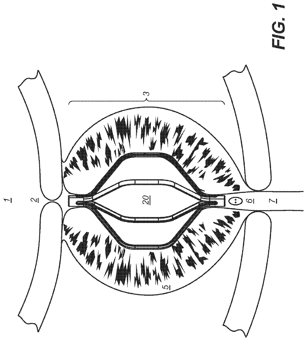
[0035]Accordingly, the method includes visualization of the prostate lobes and respective apices during implantation and orientation of the implant using the delivery system to specifically engage portions of prostate tissue by the device to place the implant into the desired configuration. The ability to incrementally deploy the implant via manipulation of the delivery system allows precise placement and orientation of the implant relative to all of the physiological structures along the length of the urethra within the transition (or T)-zone of the prostate and preferably distal to the bladder neck without obstructing the verumontanum. The method also includes the deployment of a plurality of implants selected and sized for the physiological condition of a particular BPH patient, including, the selective deployment of dissimilar embodiments of the invention as described herein and in the accompanying Figures.

Problems solved by technology

Combined with a tendency of the bladder wall to become thicker and weaker, BPH patients lose the ability to completely empty the bladder.
Urethral narrowing and urinary retention cause many of the problems experienced by BPH patients.
While these drugs provide some relief from BPH, they have unavoidable side effects and do not offer a complete solution for many BPH patients.
Other BPH patients do not experience significant alleviation of symptoms, and many find the requirement for daily medication both bothersome and costly.
However, these traditional surgical approaches to the treatment of BPH are invasive, non-reversible, and have significant drawbacks including the placement of a temporary catheter for a few months, risk of infection, loss of sexual function, urinary incontinence, and restenosis—wherein recurring hyperplasia of cells in the prostate regrow to cause a recurrence of the narrowing of the urethra opening and also a recurrence of the LUTS symptoms described above.
Although removing prostatic tissue relieves some BPH symptoms, tissue removal by traditional surgical approaches is irreversible and any adverse effects of the surgery may afflict the patient for life or affect the patients' quality of life.
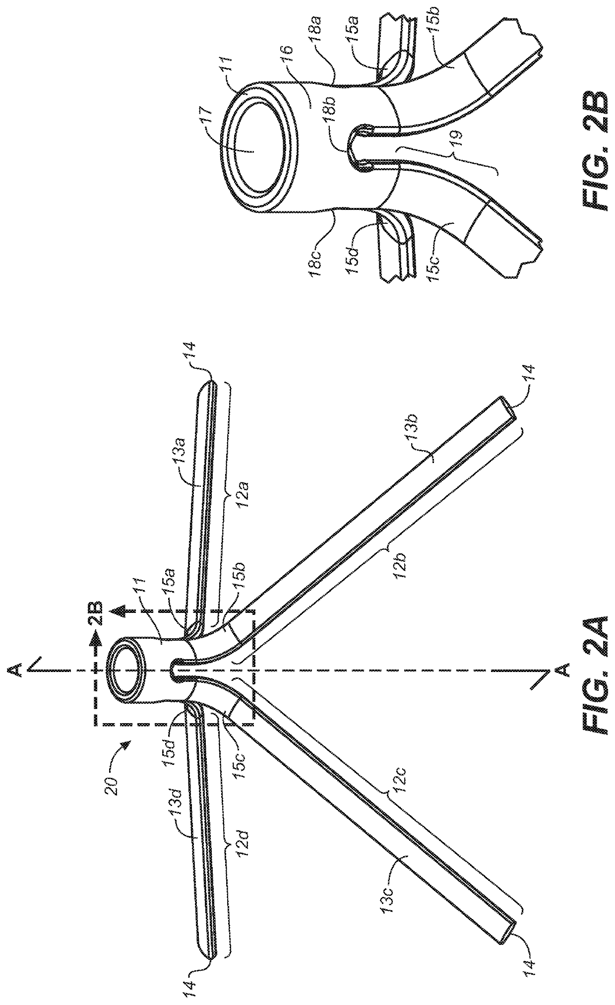
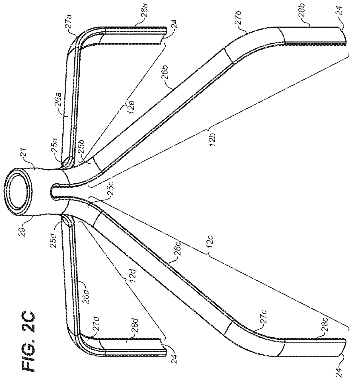
Moreover, surgical approaches are associated with the inherent risks from the surgery itself, risk recurrence from the regrowth of removed prostatic tissue, and, depending on the extent of the disease and the particular surgical approach necessary for an individual patient, can require recovery periods as long as 3 to 6 weeks.
Then, super-heated water flows through the catheter into the treatment balloon, which heats and destroys the surrounding prostate tissue.
Ultrasound energy waves from the probe heat and destroy enlarged prostate tissue.
While these less invasive techniques are generally less traumatic than traditional surgery, each destroys prostatic tissue and is irreversible.
While such mesh-style apparatus does not destroy prostate tissue, they have a tendency to migrate within the urethra and into the urinary bladder.
These designs have the drawback of disrupting the epithelial layer of cells on the interior of the urethra, causing injury to the urethral wall and risking bleeding, infection, hematuria, abnormal tissue growth, formation of stones or other trauma around the point of attachment of the implant to the urethral wall.
Mesh-like urethral stent designs also have the disadvantage of having a high implant surface area relative to the prostatic tissue area over which they apply their expansion or retraction force.
Too high a retraction force may cause significant pain to the patient and damage the urethral wall.
Higher implant surface area also increases the probability for encrustation and stone formation on the surface of the implant over time, thereby causing either urethral narrowing or structural degradation of the implant.
Inserting the rigid sheaths and endoscopes (or cystoscopes) through the penis into the prostatic urethra could be very painful.
Also, the delivery and deployment of the implants described above, may be challenging since they could obstruct direct visualization of the urethra during device placement.

Method used


Image

  • Implantable devices and methods to treat benign prostate hyperplasia (BPH) and associated lower urinary tract symptoms (LUTS)
  • Implantable devices and methods to treat benign prostate hyperplasia (BPH) and associated lower urinary tract symptoms (LUTS)
  • Implantable devices and methods to treat benign prostate hyperplasia (BPH) and associated lower urinary tract symptoms (LUTS)

Examples


Embodiment Construction

[0048]Definitions: The terms “therapeutically effective displacement” or “therapeutically effective retraction” or “therapeutically effective expansion”, are used interchangeably herein and refer to an amount of displacement of prostatic tissue proximate to a restricted area of a urethra sufficient to increase the urethral lumen and treat, ameliorate, or prevent the symptoms of benign prostatic hyperplasia (BPH) or comorbid diseases or conditions, including lower urinary tract symptoms (LUTS), wherein the displacement of prostatic tissues exhibits a detectable therapeutic, prophylactic, or inhibitory effect. The effect can be detected by, for example, an improvement in clinical condition, or reduction in symptoms or absence of co-morbidities. Examples of clinical measures include a decrease in the international prostate symptom score (IPSS), reduction in post-void residual (PVR) volume of urine in the bladder after relief or increase in the maximum urinary flow rate (Qmax) or improv...


PUM


Abstract

The invention is devices and methods to treat benign prostatic hyperplasia (BPH) and associated lower urinary tract symptoms infections (LUTS). The devices are intra-urethral implants placed in a patient in need thereof by minimally invasive procedures, preferably under local anesthesia in an office environment. The devices are sized and designed for atraumatic insertion and expansion within the urethra to engage and retract enlarged prostatic tissue proximate to the urethra that is leading to adverse symptoms associated with BPH. The methods include steps to deploy the implant devices of the invention using a delivery system of the invention and at target prostatic tissue that is visualized during the procedure and yields a reduction in the symptoms of BPH.

Description

CROSS REFERENCE TO RELATED APPLICATIONS[0001]This application claims priority to U.S. Provisional Application 62 / 502,056, filed May 5, 2017. The priority of this application is expressly claimed, and the disclosure is hereby incorporated by reference in its entirety.BACKGROUND OF THE INVENTION[0002]The prostate is a walnut-shaped gland that wraps around the urethra through which urine is expelled from the bladder and plays a crucial role in the reproductive system of men. Although the gland starts out small, it tends to enlarge as a man ages. An excessively enlarged prostate results in a disease known as benign prostatic hyperplasia (BPH). Benign prostatic hyperplasia (BPH) refers to the abnormal, but non-malignant (non-cancerous) growth of the prostate observed very commonly in aging men. BPH is a chronic condition and is associated with the development of urinary outflow obstruction in the prostatic urethra. It also causes a range of disorders referred to collectively as Lower Uri...

Claims


Application Information

Patent Timeline
Patent Type & Authority Patents(United States)
IPC IPC(8): A61F2/04A61F2/90A61B17/00A61B17/3207A61F2/962A61F2/86A61M27/00A61B17/3209A61F2/95
CPCA61F2/90A61B17/00234A61B17/320725A61F2/04A61F2/86A61F2/962A61B17/3209A61B2017/00274A61B2017/32096A61F2002/047A61F2002/9528A61F2220/0008A61F2230/001A61F2230/0073A61M27/008
Inventor HUANG, MARKCHANG, KENNETH CHIH-PINGJEN, JIMMYJANG, YUE-TEHHSU, THOMASHSU, SENZANVENKATESWARA-RAO, KONDAPAVULUR T.
Owner PRODEON MEDICAL CORP
Learn More