Determining endotracheal tube placement using acoustic reflectometry

a technology of tracheal tube placement and acoustic reflectometry, which is applied in the field of intubation, can solve the problems of inability to direct visualization, inability to detect expired carbon dioxide, and inability to have fiberoptic bronchoscopy

Inactive Publication Date: 2003-02-20
ALFRED E MANN INST FOR BIOMEDICAL ENG AT THE UNIV OF SOUTHERN CALIFORNIA
View PDF0 Cites 67 Cited by
  • Summary
  • Abstract
  • Description
  • Claims
  • Application Information

AI Technical Summary

Problems solved by technology

Direct visualization may not always be possible because of certain patient conditions, particularly in difficult airway patients.
Fiberoptic bronchoscopy may not always be available, especially in emergency settings outside the operating room.
Detection of expired carbon dioxide may not be possible in patients such as those with cardiopulmonary arrest where there is no characteristic color change in the calorimetric device and/or a wave form is absent in a capnograph or spectrometer trace.
However, this device is sometimes slow, requiring up to thirty seconds for a determination.
It is often not usable in infants less than 1 year of age, having a failure rate of approximately 25%.
It can also generate false results when there is an airway obstruction, such as a foreign body, asthma, or a mediastinal mass compressing the trachea.
However, even these devices may present spurious results.
It is the critical failure to recognize an esophageal intubation that continues to

Method used


Image

  • Determining endotracheal tube placement using acoustic reflectometry
  • Determining endotracheal tube placement using acoustic reflectometry
  • Determining endotracheal tube placement using acoustic reflectometry

Examples


Embodiment Construction

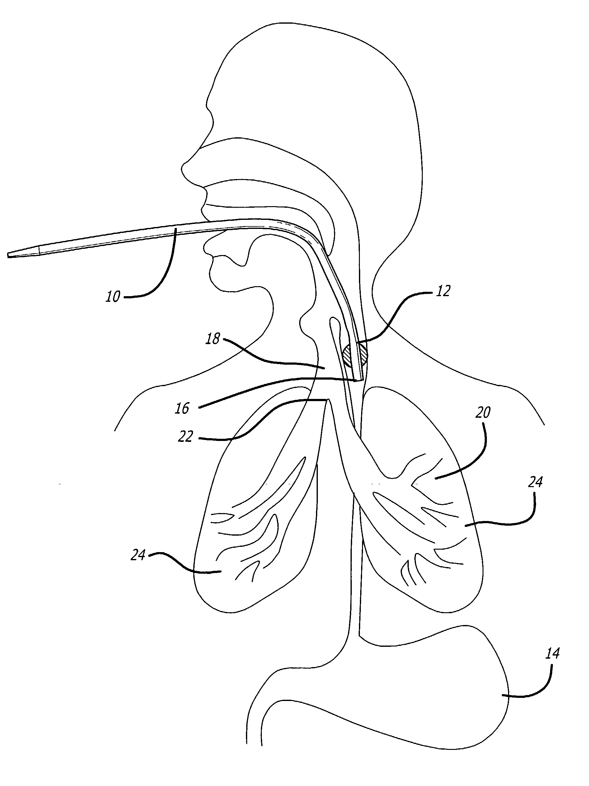
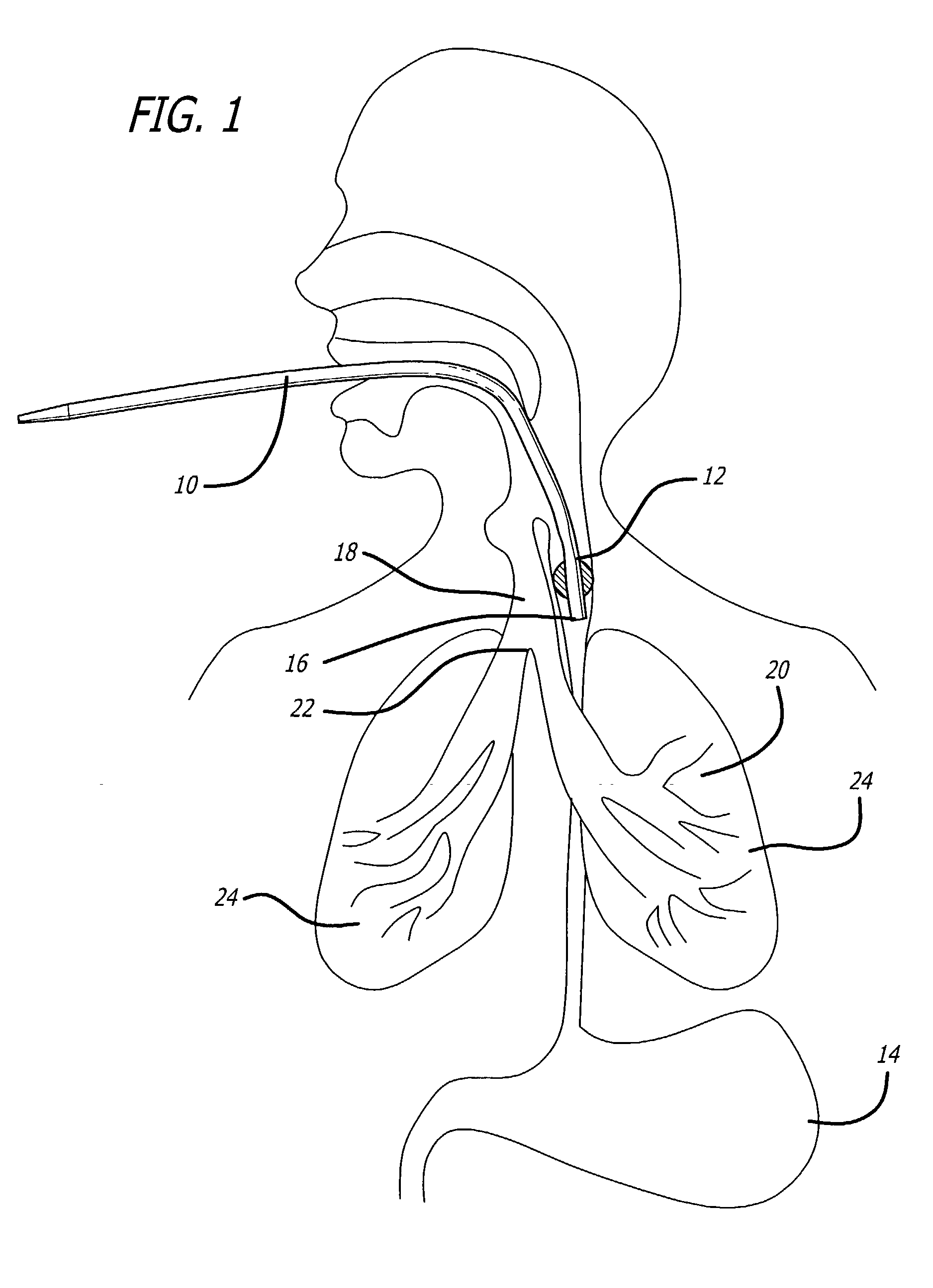
[0048] FIGS. 1-3 are side elevational views showing an endotracheal tube ("ETT") 10 inserted into different anatomical sites in a patient.

[0049] FIG. 1 is a side elevational view showing the ETT improperly inserted into the patient's esophagus 12 which, of course, leads to the patient's stomach 14. The distal end 16 of the ETT 10is shown in the esophagus 12 just below the entranceways to the esophagus 12 and trachea 18.

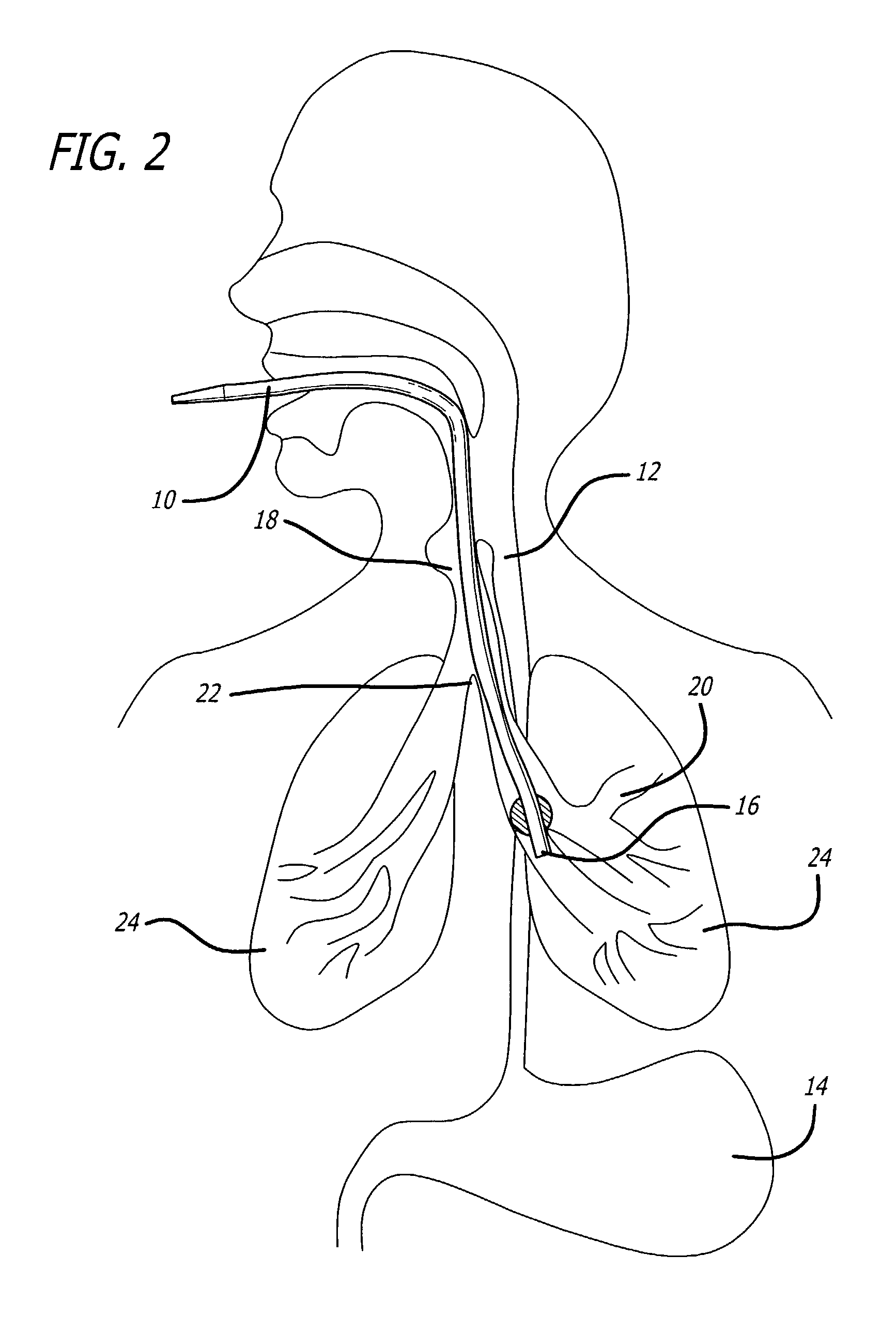
[0050] FIG. 2 is a side elevational view showing the ETT improperly inserted into a patient's bronchus 20. An endobronchial intubation such as this occurs when the distal end 16 of an ETT 10 is advanced through a patient's trachea 18, and advanced further beyond the carina 22, into the left (as shown) or right (not shown) mainstem bronchus 20. In such a situation, ventilation of only one of a patient's lungs 24 is possible.

[0051] FIG.3 is a side elevational view showing an ETT 10 properly inserted into a patient's trachea 18. The distal end 16 of the ETT 10 is positio...


PUM


Abstract

Determining the placement of an endotracheal tube in a patient. The invention evaluates discontinuities in the medium surrounding the endotracheal tube, such as the airway, as a function of distance past an end of the endotracheal tube. Using a loudspeaker to generate sound waves, the sound waves propagate through a coiled wavetube, a connecting adapter, and an endotracheal tube, into the area of interest. With a processing system, reflected sound waves which return from the cavity back to a microphone within the wavetube are analyzed and an area-distance curve of the area in interest is constructed.

Description

[0001] This application claims priority to and incorporates by reference U.S. Provisional Application No. 60 / 312,283, filed Aug. 14, 2001, entitled "Miniature, Portable Acoustic Reflectometer."[0002] 1. Field of the Invention[0003] The invention relates to intubations and, more particularly, determining whether an endotracheal tube ("ETT") has been correctly placed and positioned.[0004] 2. Description of Related Art[0005] In the pre-hospital setting, the incidence of unrecognized esophageal intubations varies between 1.8-2.0%. Current methods that assist physicians in correctly performing an endotracheal intubation consist of direct visualization of the passage of the ETT through the vocal cords, fiberoptic bronchoscopy, and detection of expired carbon dioxide. Direct visualization may not always be possible because of certain patient conditions, particularly in difficult airway patients. Fiberoptic bronchoscopy may not always be available, especially in emergency settings outside t...

Claims


Application Information

Patent Timeline
IPC IPC(8): A61M16/04
CPCA61M16/0488A61M2209/02A61M2205/3375A61M16/0411
Inventor RAPHAEL, DAVID T.
Owner ALFRED E MANN INST FOR BIOMEDICAL ENG AT THE UNIV OF SOUTHERN CALIFORNIA
Why Eureka
Try Eureka