

Method for the Detection of Microorganisms by Fiber Optics
a technology of fiber optics and microorganisms, applied in the field of microorganism detection by fiber optics, can solve the problems of other types of microorganisms being intrinsically dangerous, microorganisms not serving any other practical purpose, and microorganisms becoming dangerous, etc., to achieve the effect of easy incorporation, easy operation and miniaturization
- Summary
- Abstract
- Description
- Claims
- Application Information
AI Technical Summary
Benefits of technology
Problems solved by technology
Method used
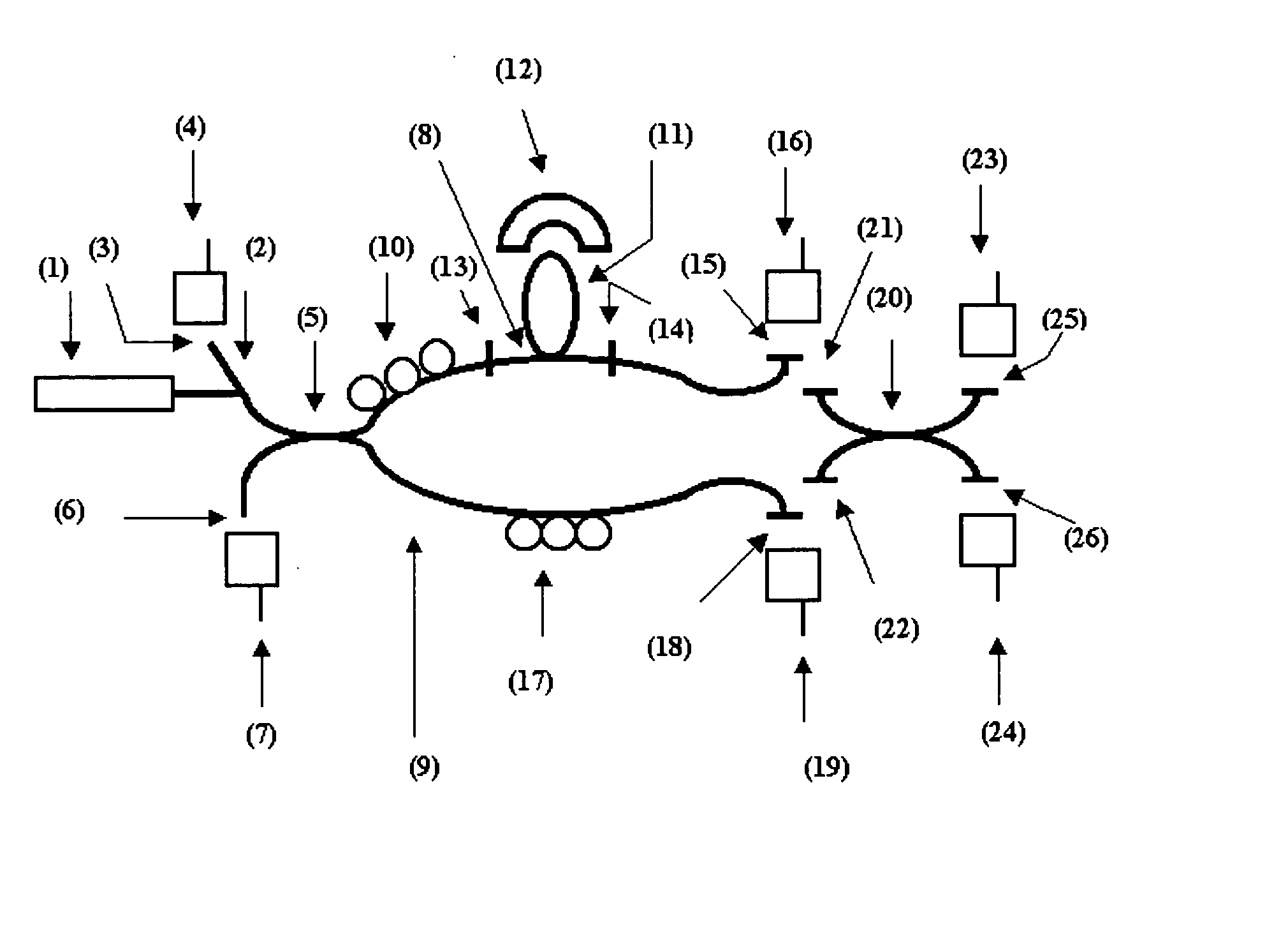
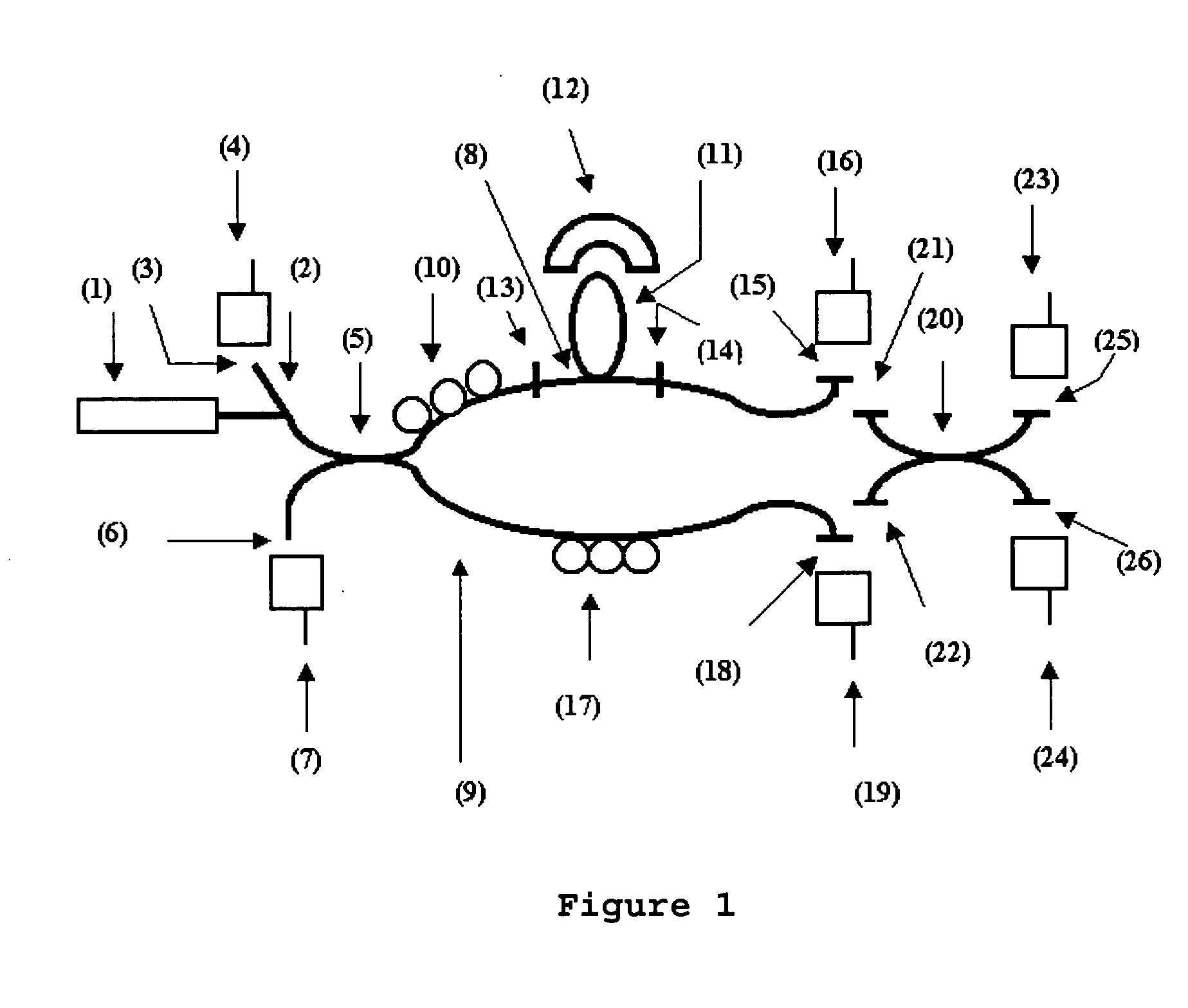
Image

Examples
example 2
Obtaining an Air Sample
[0043] An air sample is collected through the technique known to experts in the field as impaction-on-a-gel employing Merck MAS-100 equipment. In this manner, a sample of the ambient air was aspirated, through perforated plates, with the aid of a vacuum pump (with a volumetric flow rate of 100 litres per minute) for 30 seconds. The resulting air stream that carries particles with diameters inferior to 10 .mu.m, was directed to the agar surface of a Petri dish. Eighteen collections were undertaken, during six random days, three collections per day, during two weeks. During the three first days, a specific culture medium for Staphylococcus aureus was used, and during the three remaining days, a specific medium for S. pneumoniae was used.
example 3
Growth of Microorganisms in a Selective Culture Medium
[0044] The culture medium used for Staphylococcus aureus resistant to methicillin (MRSA) obtained in Example 2 was the Baird-Parker Agar (Difco Laboratories-Difco 0768-17-3) at an incubation temperature of 35.degree. C. For S. pneumoniae, the culture medium used was Trypticase soya agar (Difco Laboratories, Detroit, Mich.--Difco 0026-17-1) supplemented with sheep blood at 5% at an incubation temperature of 35.degree. C. In both the culture media glycerol at 0,2% was added, so as to avoid drying out the culture during the tests. For the growth of E. coli 0157:H7, the selective culture medium employed was the MacConkey sorbitol agar base (Difco Laboratories-Difco 0079-17-7), at an incubation temperature of 35.degree. C.
[0045] Many tests were performed so as to calibrate the biosensor for its selectivity, using different initial values of bacteria (No). E. coli 0157:H7 available commercially in lyophilized form was reconstituted wit...
example 4
Correlation of the Growth with the Estimated Number of Bacteria
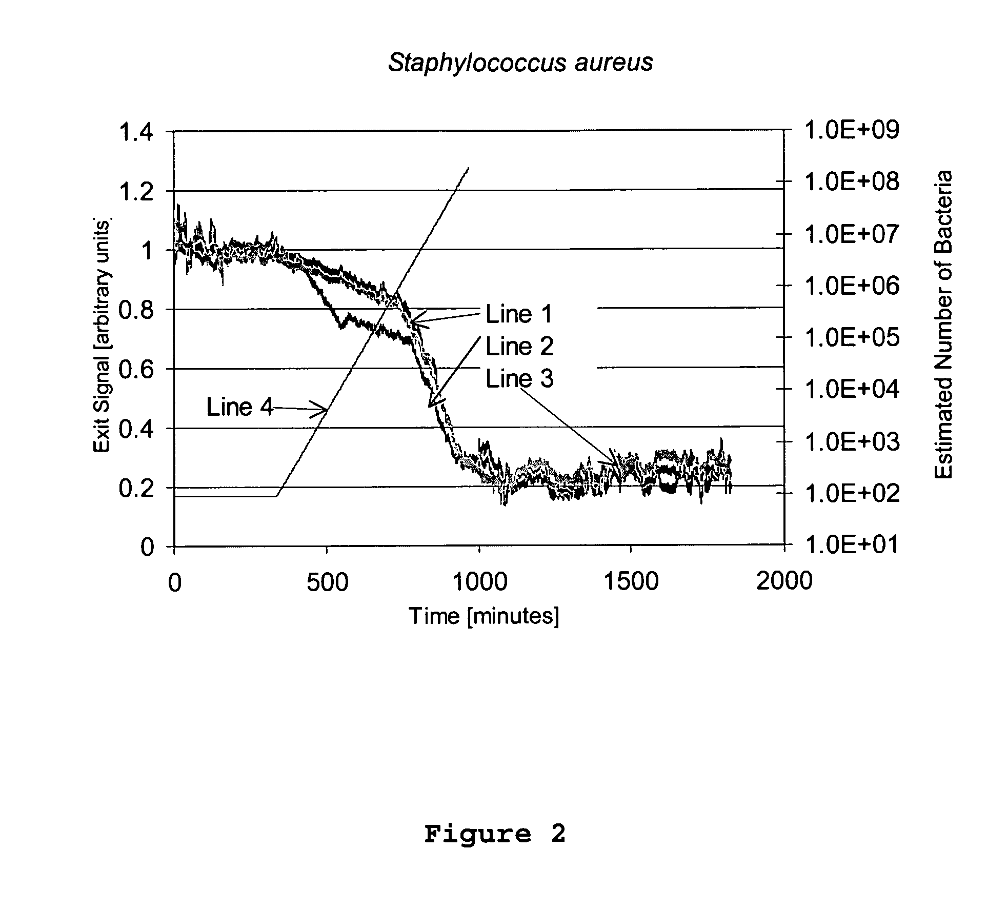
[0046] FIGS. 2 and 3 present, respectively, the optical signal of the sensor for Staphylococcus aureus resistant to methicillin and S. pneumoniae. Lines 1, 2 and 3 represent the output signal for samples 1, 2 and 3, respectively. Line 4 correlates the growth with the estimated number of bacteria, in accordance with the following formula:
N(t)=N.sub.0 2.sup.t / GT
[0047] where t is the time in minutes, N is the total number of bacteria in the time t, N.sub.0 is the initial number of bacteria (equal to 94 for Staphylococcus aureus resistant to methicillin (MRSA) and 91 for S. pneumoniae) and GT is the time of generation (equal to 30 minutes for MRSA and 48 minutes for S. pneumoniae).
[0048] Observing FIGS. 2 and 3, one notes the presence of three distinct phases: the first is a plateau where no response is detected (Lag phase). The result for Staphylococcus aureus resistant to methicillin and S. pneumoniae demonstrate a lag pha...
PUM
| Property | Measurement | Unit |
|---|---|---|
| thickness | aaaaa | aaaaa |
| thickness | aaaaa | aaaaa |
| diameter | aaaaa | aaaaa |
Abstract
Description
Claims
Application Information