

Pre-operative medical planning system and method for use thereof
a medical planning and preoperative technology, applied in image data processing, analogue and hybrid computing, sensors, etc., can solve the problems of insufficient challenge of digital radiology, difficulty in saving or printing, and use as a rough guide, so as to reduce fractures and prevent incorrect positioning of fixation elements
- Summary
- Abstract
- Description
- Claims
- Application Information
AI Technical Summary
Benefits of technology
Problems solved by technology
Method used
Image

Examples
Embodiment Construction
[0036] The following description is provided, alongside all chapters of the present invention, so as to enable any person skilled in the art to make use of said invention and sets forth the best modes contemplated by the inventor of carrying out this invention. Various modifications, however, will remain apparent to those skilled in the art, since the generic principles of the present invention have been defined specifically to provide methods and apparatus for pre-operative planning of orthopedic surgical procedures.
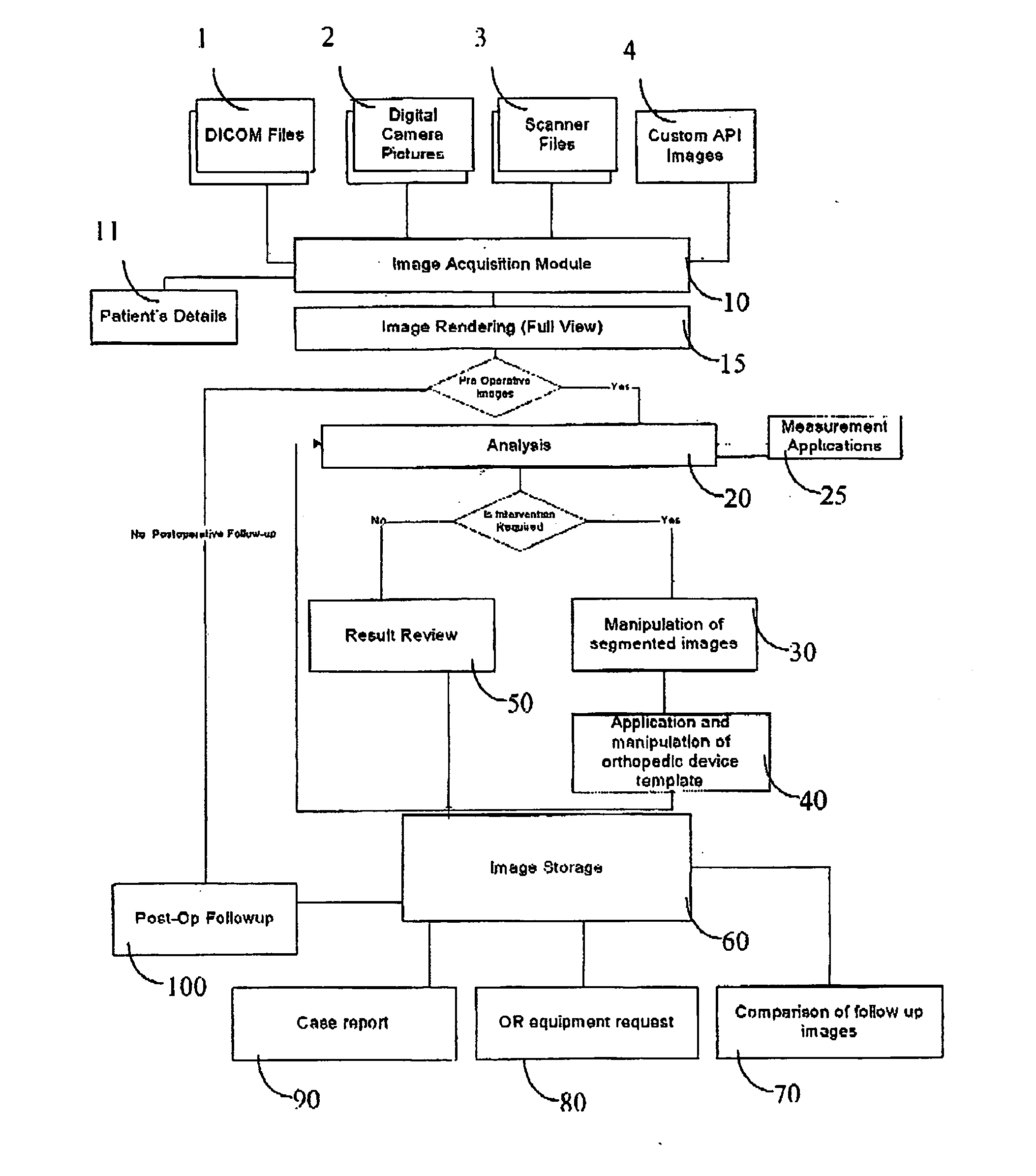
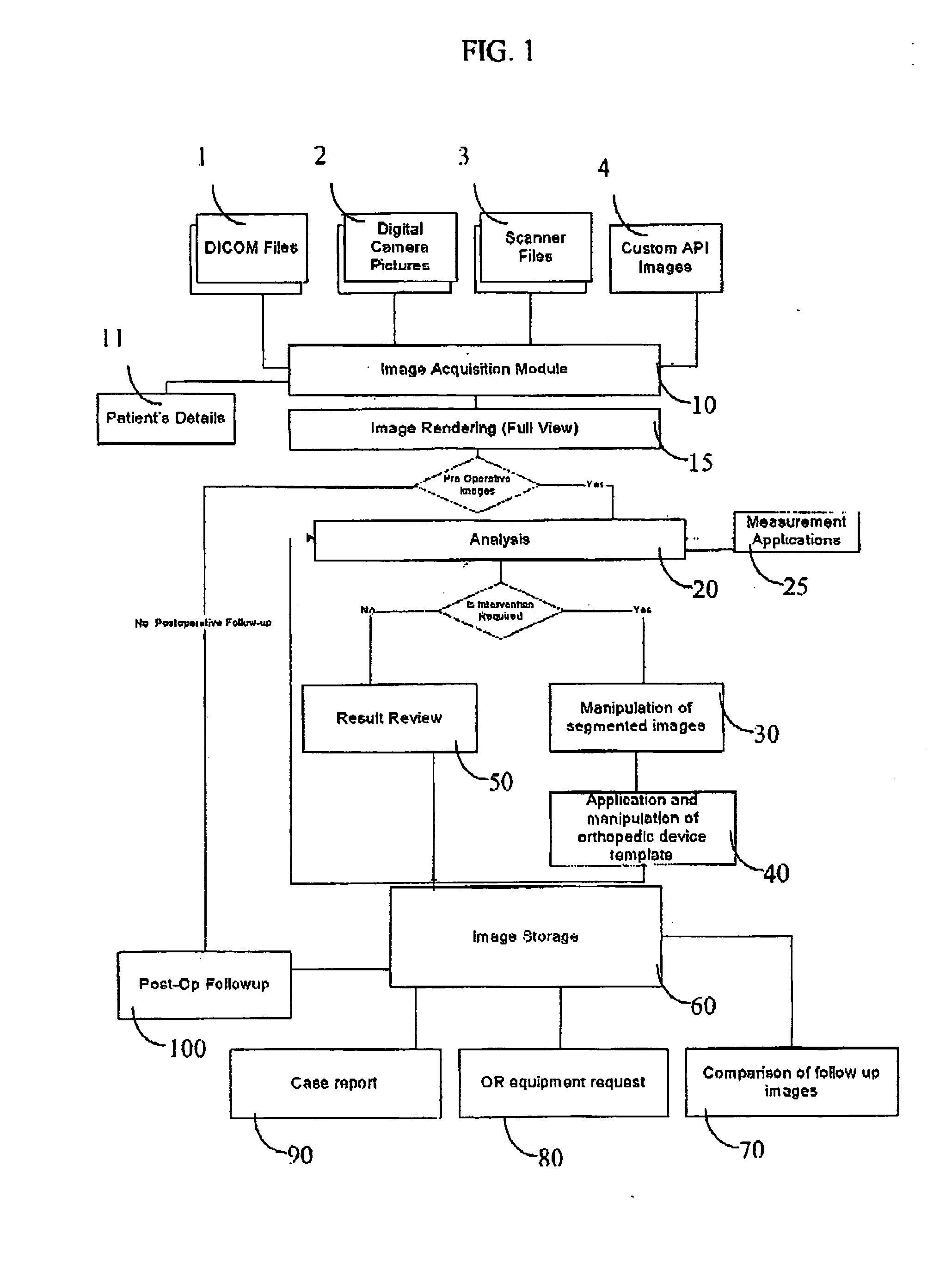
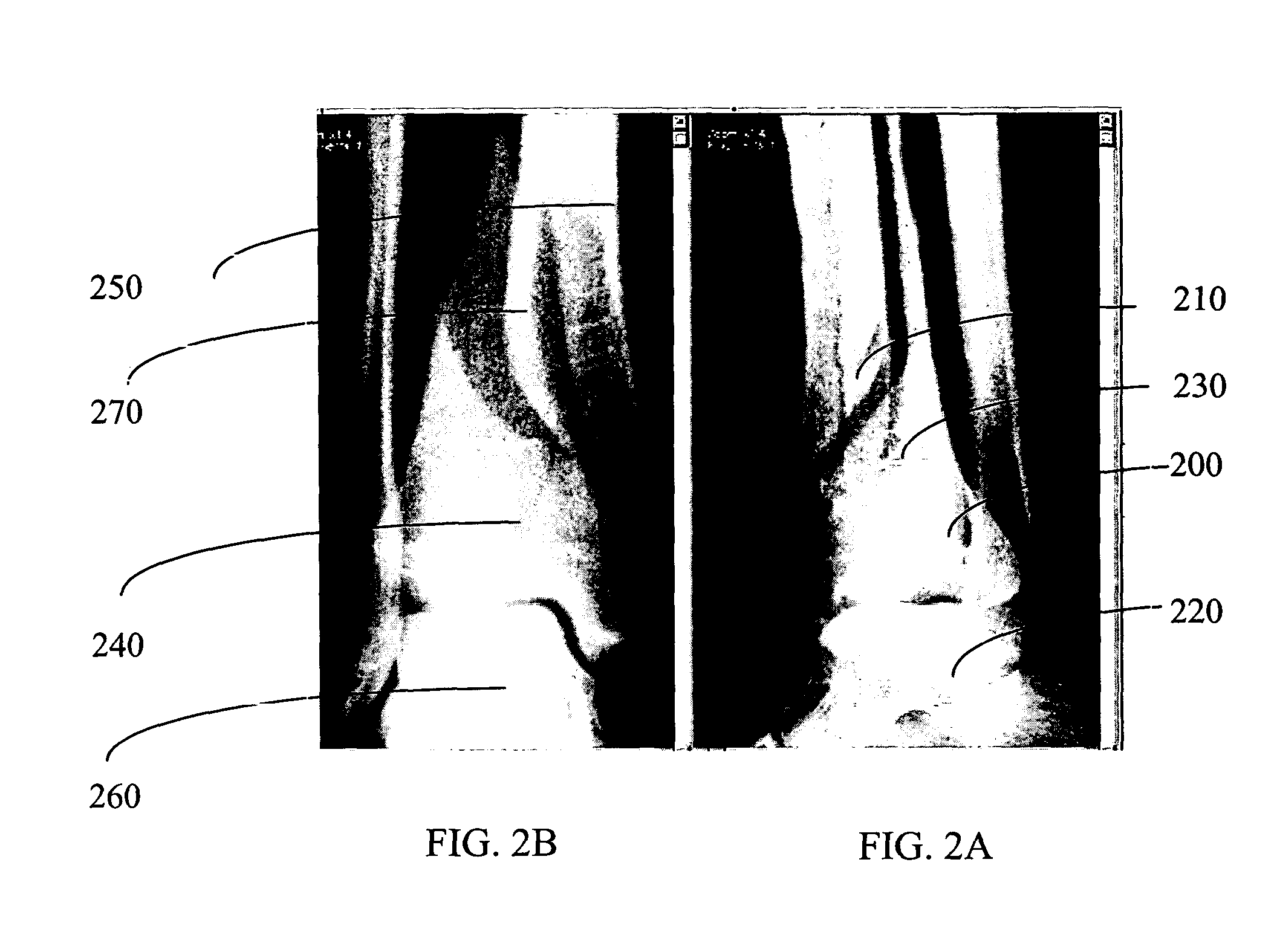
[0037] The present invention is directed to a method for pre-planning and simulating of orthopaedic surgical procedures using medical images. The method comprises the following steps: (a) obtaining and displaying the medical images; (b) segmenting anatomical structure segments in the medical images; and (c) planning the desired result of the orthopaedic surgical procedure so output images are produced, wherein the obtained output images comprise features selected from ...
PUM
 Abstract
Description
Claims
Application Information