Apparatus and methods for mapping and ablation in electrophysiology procedures

a technology of electrophysiology and apparatus, applied in the field of medical devices, can solve the problems of premature death of people, decreased amount of blood pumped by the heart, and complex organ of human hear

Inactive Publication Date: 2005-03-24
BOSTON SCI SCIMED INC
View PDF63 Cites 109 Cited by
  • Summary
  • Abstract
  • Description
  • Claims
  • Application Information

AI Technical Summary

Problems solved by technology

The human heart is a very complex organ, which relies on both muscle contraction and electrical impulses to function properly.
When the chambers contract at improper times, the amount of blood pumped by the heart decreases, which can result in premature death of the person.
Though mapping and ablation of these triggers appears to be curative in patients with paroxysmal atrial fibrillation, there are a number of limitations to ablating focal triggers via mapping and ablating the earliest site of activation with a “point” radiofrequency lesion.
Such a technique is relatively involved, and requires significant skill and attention from the clinician performing the procedures.

Method used


Image

  • Apparatus and methods for mapping and ablation in electrophysiology procedures
  • Apparatus and methods for mapping and ablation in electrophysiology procedures
  • Apparatus and methods for mapping and ablation in electrophysiology procedures

Examples


Embodiment Construction

[0037] System Overview

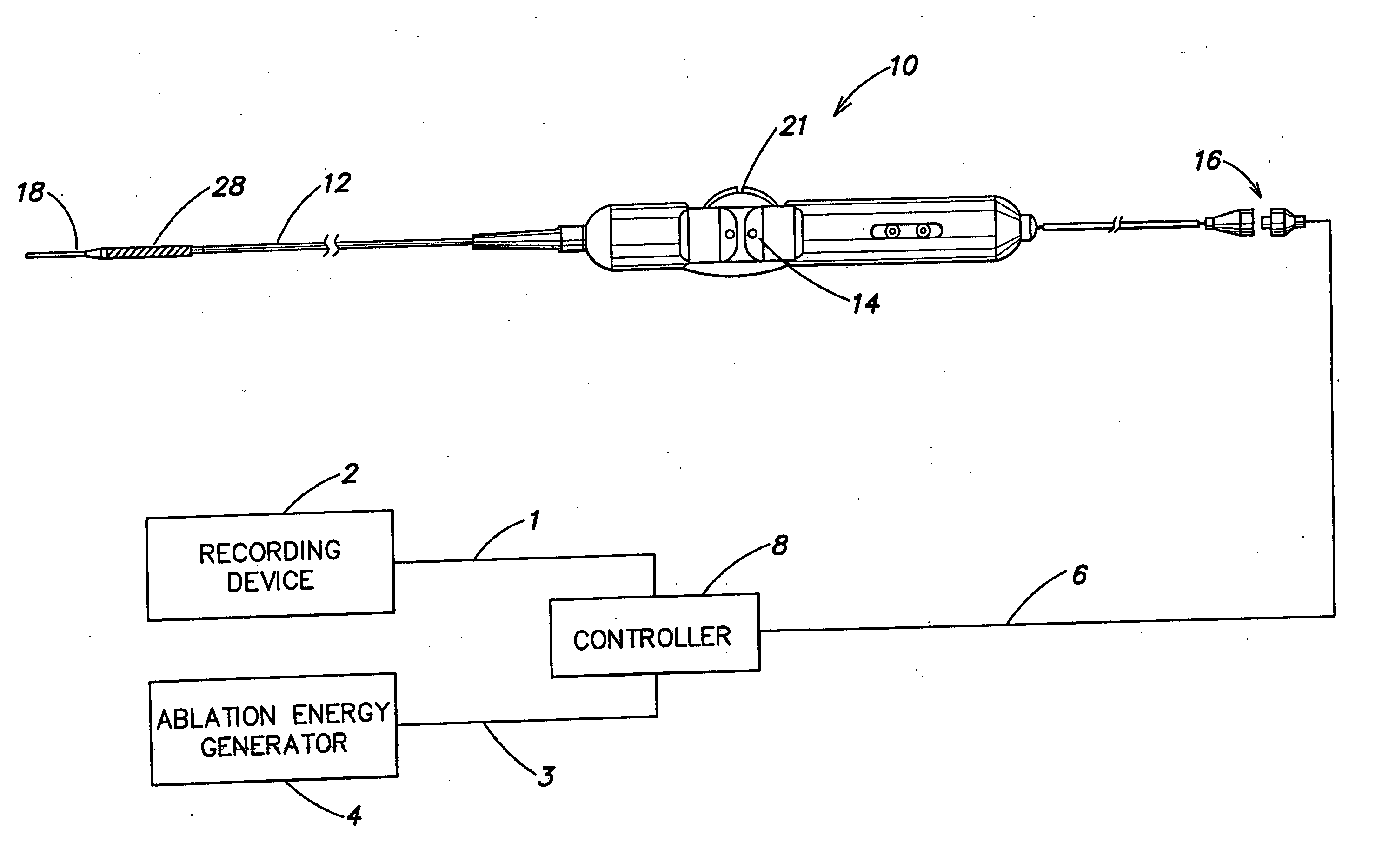
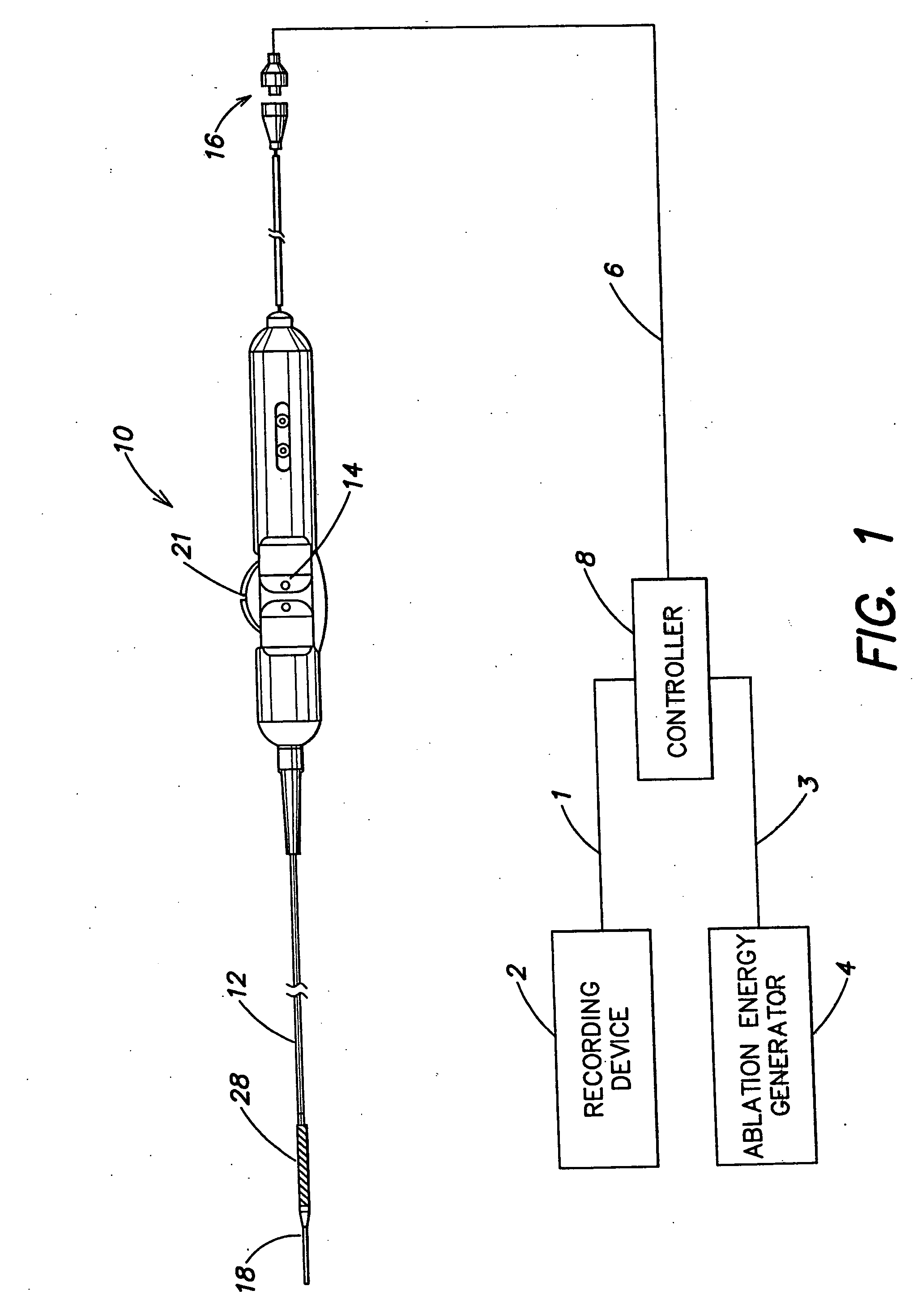
[0038] Reference is now made to FIG. 1, which figure illustrates an overview of a mapping and ablation catheter system in accordance with the present invention. The system includes a catheter 10 having a shaft portion 12, a control handle 14, and a connector portion 16. A controller 8 is connected to connector portion 16 via cable 6. Ablation energy generator 4 may be connected to controller 8 via cable 3. A recording device 2 may be connected to controller 8 via cable 1. When used in an ablation application, controller 8 is used to control ablation energy provided by ablation energy generator 4 to catheter 10. When used in a mapping application, controller 8 is used to process signals coming from catheter 10 and to provide these signals to recording device 2. Although illustrated as separate devices, recording device 2, ablation energy generator 4, and controller 8 could be incorporated into a single device. In one embodiment, controller 8 may be a QUADRAPULS...


PUM


Abstract

An electrophysiology catheter and method of use for mapping and ablation procedures. The catheter includes a braided conductive member at its distal end that can be radially expanded. The catheter can be used in endocardial and epicardial mapping and ablation procedures.

Description

CROSS REFERENCE TO RELATED APPLICATIONS [0001] This application is a divisional of U.S. application Ser. No. 09 / 845,022, filed Apr. 27, 2001 entitled APPARATUS AND METHODS FOR MAPPING AND ABLATION IN ELECTROPHYSIOLOGY PROCEDURES, which is hereby incorporated herein by reference in its entirety, and which, in turn, claims the benefit of U.S. Provisional Application Ser. No. 60 / 261,015 entitled HIGH DENSITY MAPPING AND ABLATION CATHETER AND METHOD OF USE, filed Jan. 11, 2001; U.S. Provisional Application Ser. No. 60 / 204,457 entitled METHOD FOR CREATING ANNULAR EPICARDIAL LESIONS AT THE OSTIA OF THE PULMONARY VEINS, filed on May 16, 2000; U.S. Provisional Application Ser. No. 60 / 204,482 entitled METHOD AND DEVICE FOR CREATING ANNULAR ENDOCARDIAL LESIONS AT THE OSTIA OF THE PULMONARY VEINS, filed May 16, 2000; and U.S. Provisional Application Ser. No. 60 / 201,445 entitled TRANSMURAL CIRCUMFERENTIAL LESIONS MADE AT CANINE PV OSTIUM BY EXPANDABLE MESH ELECTRODES IN VIVO, filed May 3, 2000,...

Claims


Application Information

Patent Timeline
Patent Type & Authority Applications(United States)
IPC IPC(8): A61B5/01A61B5/296A61B18/00A61B18/02A61B18/12A61B18/14A61B18/18
CPCA61B2018/00577A61B2018/00029A61B2018/1497A61B2018/00946A61B2018/00267A61B2018/0016A61B2018/00821A61B2018/00214A61B2018/00083A61B18/1492A61B2018/00196A61B2017/003A61B2018/00797
Inventor COLLINS, RUSSELL F.FALWELL, GARY S.BENE, ERIC A.BURNS, STEVEN J.COLLINS, DENYSE M.GIBSON, CHARLES A.HE, DING SHENGLECLAIR, PAUL E.PATTERSON, DONALD F.SAGON, STEPHEN W.JAIS, PIERRE
Owner BOSTON SCI SCIMED INC
Why Eureka
Try Eureka