Method and apparatus for indicating an encountered obstacle during insertion of a medical device

a medical device and anatomical technology, applied in the field of medical devices, can solve the problems of non-radiographic alternatives, inventions that do not image the internal anatomy of patients, and carry ionizing radiation risks for both patients and caregivers

Inactive Publication Date: 2005-04-07
LUCENT MEDICAL SYST
View PDF4 Cites 1251 Cited by
  • Summary
  • Abstract
  • Description
  • Claims
  • Application Information

AI Technical Summary

Problems solved by technology

This imaging necessitates the use of expensive equipment, facilities, and personnel, and radiographic imaging also carries ionizing radiation risks to both the patient and caregivers.
However, these inventions do not image a patient's internal anatomy, and the caregiver relies upon the correspondence between external anatomical landmarks and the magnet's displayed location underneath those landmarks to correctly place the medical device.
For example, a cardiac catheter may be unintentionally pushed up against the wall of the right atrium, which could lead to cardiac injury or even puncture, resulting in death.
Although radiographic imaging can show this dangerous situation to the caregiver, the non-radiographic alternatives, at present, do not.

Method used


Image

  • Method and apparatus for indicating an encountered obstacle during insertion of a medical device
  • Method and apparatus for indicating an encountered obstacle during insertion of a medical device
  • Method and apparatus for indicating an encountered obstacle during insertion of a medical device

Examples


Embodiment Construction

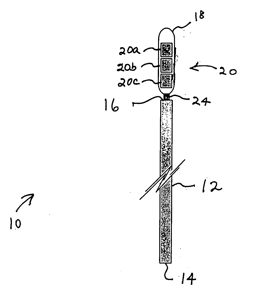
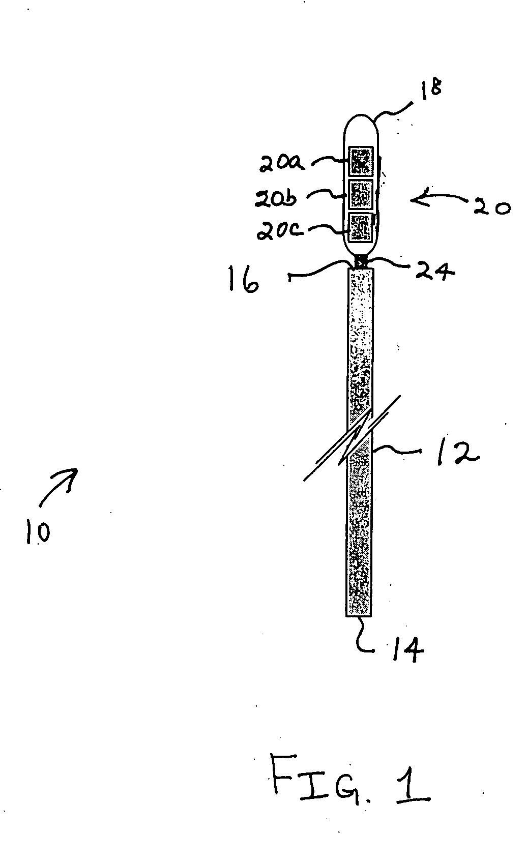
[0020] As will be discussed in greater detail herein, a method and apparatus disclosed herein can indicate internal, anatomical obstacles encountered during medical device insertion by allowing the inserted medical device's tracked element, when attached to the distal end of the medical device, to easily bend at an angle to the direction of travel.

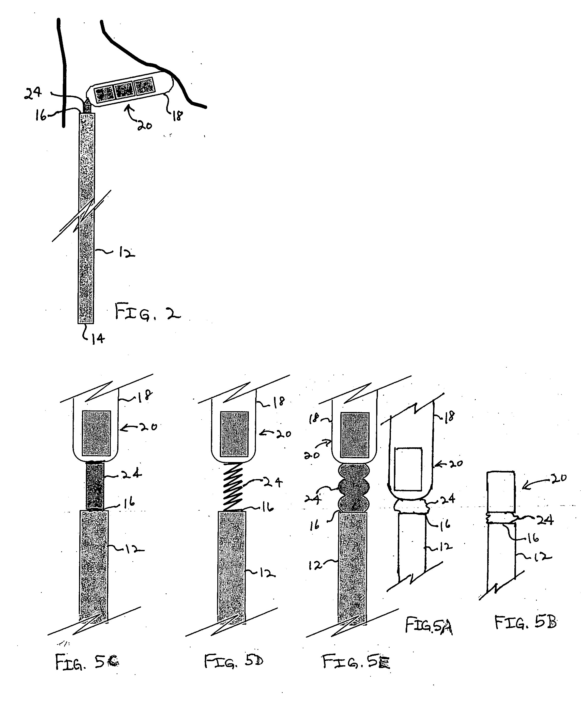
[0021] An exemplary embodiment of the present invention is illustrated in a system 10 illustrated in FIG. 1. The system 10 includes a medical elongated member 12 having a proximal end portion 14 and a distal end portion 16. The distal end portion 16 is designed for insertion into the body. For example, the elongated member 12 may be designed as a cardiac catheter with the distal end portion 16 being inserted into a desired location within the heart.

[0022] A vessel or chamber 18 is flexibly coupled to the distal end portion 16 of the elongated member 12. Contained within the chamber 18 is a location indicating element 20. In one embodimen...


PUM


Abstract

A technique for detecting and indicating an internal anatomical obstacle encountered during insertion of a medical device into the body of a patient, comprising an elongated member such as a tube, catheter, guidewire, or other device, having a location indicating element, such as a permanent magnet, flexibly coupled to its distal end, and an external detector that tracks and displays the location and orientation of the location indicating element. The flexible coupling has sufficient stiffness to maintain the orientation of the location indicating element against the forces from both gravity and flowing blood within a patient's vasculature, but allows the location indicating element to change orientation if it encounters an obstacle during insertion. The medical caregiver monitors the detector's display and determines encounters with obstacles by observing changes in the orientation of the location indicating element.

Description

BACKGROUND OF THE INVENTION [0001] 1. Field of the Invention [0002] The present invention is directed generally to medical devices and, more particularly, to a method and apparatus for detecting and indicating encounters with internal, anatomical obstacles during the insertion of a medical device in the body of a patient. [0003] 2. Description of the Related Art [0004] Many medical procedures require the placement of a catheter, tube, guidewire, or other device into the body of a patient, and these devices may be inserted into and / or through veins, arteries, the mouth, nose, or other openings. The ultimate destination of the distal tip of the inserted medical device may be at some distance from its insertion point. For example, a catheter may be inserted into a patient's femoral vein at the groin, with the goal of placing the catheter's distal end at the midpoint of the patient's superior vena cava, above the right atrium of the heart. [0005] Safe placement of such devices is often ...

Claims


Application Information

Patent Timeline
Patent Type & Authority Applications(United States)
IPC IPC(8): A61B5/06A61B19/00
CPCA61B5/06A61B5/062A61B5/065A61B19/52A61B19/54A61B2017/00876A61B2019/5483A61B2019/481A61B2019/4836A61B2019/4857A61B2019/5251A61B2019/5454A61B2019/306A61B90/36A61B90/39A61B2034/2051A61B2090/036A61B2090/08021A61B2090/0807A61B2090/0811A61B2090/3954A61B2090/3983
Inventor GOLDEN, ROBERT N.
Owner LUCENT MEDICAL SYST
Why Eureka
Try Eureka