System and method for processing specimens and images for optical tomography

a technology of optical tomography and processing system, applied in the field of imaging specimens, can solve problems such as optical system limitations, lateral smearing of images, and degrading images with degrading optical aberrations

Active Publication Date: 2005-04-21
UNIV OF WASHINGTON +1
View PDF82 Cites 142 Cited by
  • Summary
  • Abstract
  • Description
  • Claims
  • Application Information

AI Technical Summary

Problems solved by technology

There are a number of detrimental optical aberrations that degrade images including:
(a) lateral smearing of the image due to inherent optical system limitations which occurs even for thin specimens;
Focusing on such regions may be unavoidable as where

Method used


Image

  • System and method for processing specimens and images for optical tomography
  • System and method for processing specimens and images for optical tomography
  • System and method for processing specimens and images for optical tomography

Examples


Embodiment Construction

[0041] The method and apparatus of the invention is here described with reference to specific examples that are intended to be illustrative and not limiting. The method and apparatus of the invention is amenable to additional features such as matching of the refractive indices of the materials in the samples and the inclusion of microscopic barcodes to facilitate identification and tracking.

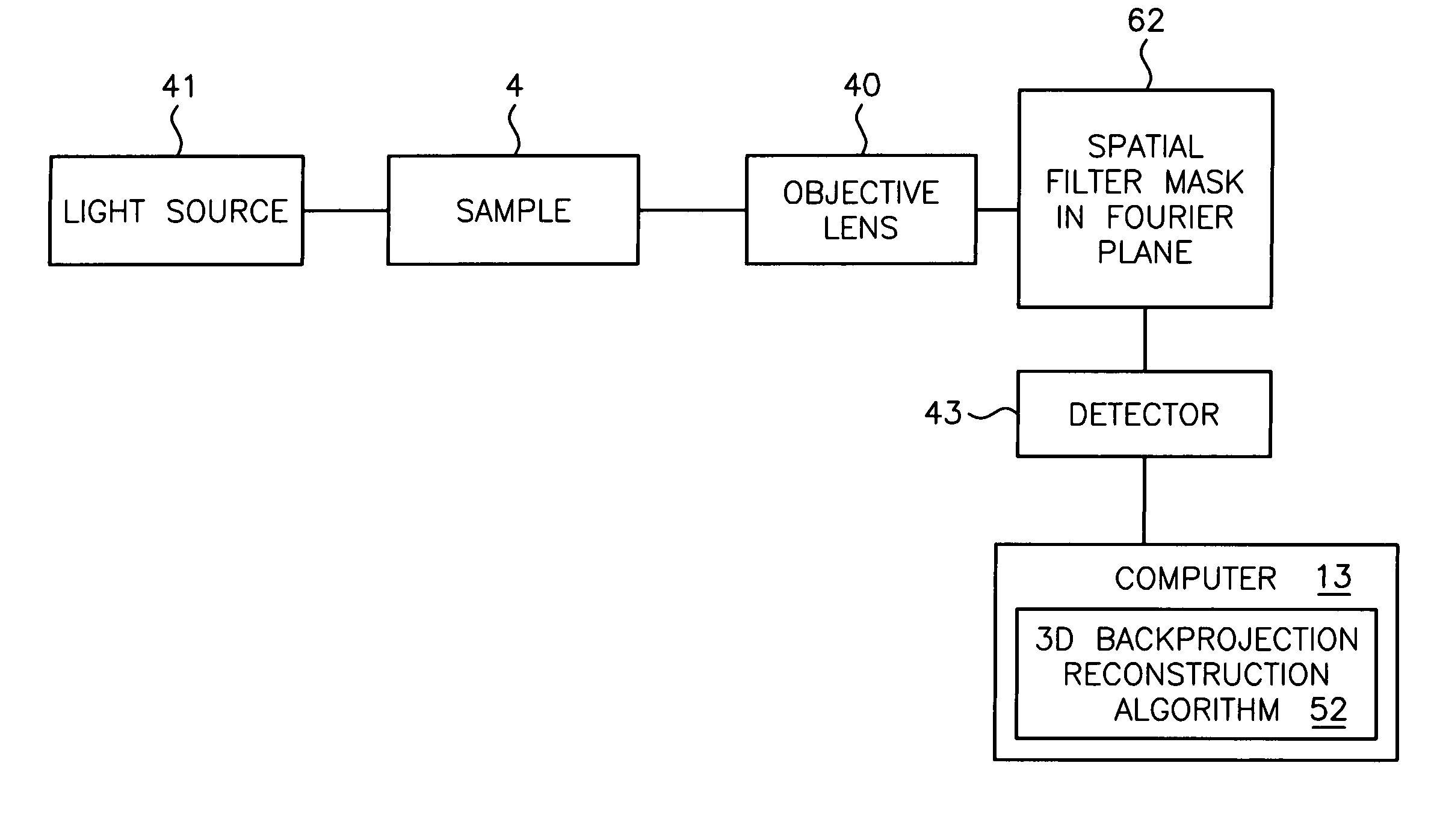
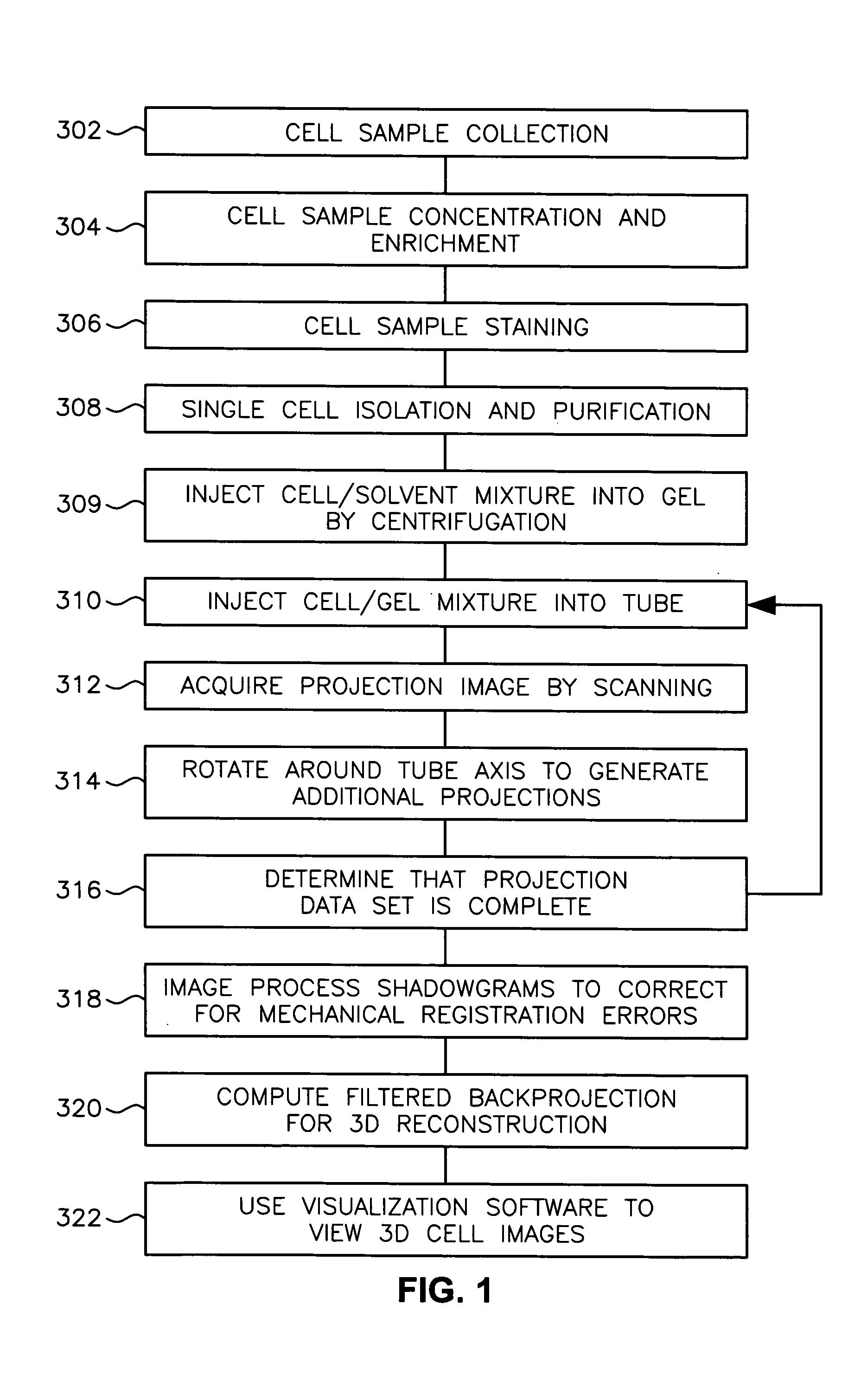
[0042] Referring now to FIG. 1, a schematic view of a high level flow diagram of a scanning system and method for scanning samples using optical tomography as contemplated by one example embodiment of the current invention is illustrated. As typically implemented, the steps may include a combination of manual operations, as in sample collection, and automated processing, as in using reconstruction algorithms or equivalent types of operations including combinations of manual and automated operations. Steps included in preparing, acquiring, reconstructing and viewing three-dimensional images begin...


PUM


Abstract

A scanning method for scanning samples of biological cells using optical tomography includes preparing, acquiring, reconstructing and viewing three-dimensional images of cell samples. Concentration and enrichment of the cell sample follows. The cell sample is stained. Cells are isolated from the cell sample and purified. A cell/solvent mixture is injected into a gel by centrifugation. A cell/gel mixture is injected into a capillary tube until a cell appears centered in a field of view using a step flow method. An optical imaging system, such as a fixed or variable motion optical tomography system acquires a projection image. The sample is rotated about a tube axis to generate additional projections. Once image acquisition is completed, the acquired shawdowgrams or image projections are corrected for errors. A computer or other equivalent processor is used to compute filtered backprojection information for 3D reconstruction.

Description

RELATED APPLICATIONS [0001] This application claims the benefit of the priority date and is a continuation-in-part of co-pending U.S. patent application Ser. No. 10 / 716,744, filed Nov. 18, 2003, of Fauver et al. entitled “METHOD AND APPARATUS OF SHADOWGRAM FORMATION FOR OPTICAL TOMOGRAPHY,” which is a continuation-in-part of co-pending U.S. patent application Ser. No. 10 / 126,026, filed Apr. 19, 2002, of Nelson entitled “VARIABLE-MOTION OPTICAL TOMOGRAPHY OF SPECIMEN PARTICLES,” the disclosures of both of which are incorporated herein by this reference.FIELD OF THE INVENTION [0002] The present invention relates generally to the field of imaging specimens and, more particularly, to imaging specimens using optical tomography. BACKGROUND OF THE INVENTION [0003] Fauver et al. teaches an optical imaging system that generates pseudoprojection images from multiple views of a specimen, such as a biological cell. Careful preparation of samples is required prior to initiating optical imaging. ...

Claims


Application Information

Patent Timeline
IPC IPC(8): A61B5/05G02B21/00G02B21/36
CPCG02B21/365G02B21/002
Inventor FAUVER, MARK E.RAHN, JOHN RICHARDSEIBEL, ERIC J.NELSON, ALAN C.
Owner UNIV OF WASHINGTON
Why Eureka
Try Eureka