Methods for therapy of neurodegenerative disease of the brain

a neurodegenerative disease and brain technology, applied in the field of brain neurodegenerative disease treatment, can solve the problems of affecting the treatment effect, and achieving limited success in direct delivery of neurotrophins through infusion into the neurocompromised brain, and achieves minimal toxicity and high transduction efficiency.

Inactive Publication Date: 2005-06-09
RGT UNIV OF CALIFORNIA
View PDF3 Cites 4 Cited by
  • Summary
  • Abstract
  • Description
  • Claims
  • Application Information

AI Technical Summary

Benefits of technology

[0009] This lentiviral based protocol for neurotrophin delivery achieves a high level of transduction efficiency, with minimal toxicity, to produce a therapeutic or preventative effect in the primate brain.

Problems solved by technology

Direct delivery of neurotrophins through infusion into the neurocompromised brain has been met with limited success and, in one instance, actually worsened the condition being treated (Kordower, et al., Ann. Neurol., 46:419-424, 1999 [symptoms of PD worsened following infusion of glial cell-derived neurotrophic factor]).

Method used


Image

  • Methods for therapy of neurodegenerative disease of the brain
  • Methods for therapy of neurodegenerative disease of the brain
  • Methods for therapy of neurodegenerative disease of the brain

Examples


example i

Construction of GDNF Expressing Lentiviral Vector

[0053] The cDNA coding for a nuclear-localized β-galactosidase (LacZ) and the human GDNF containing a Kozak consensus sequence (a 636-bp fragment: position 1 to 151 and 1 to 485; GenBank accession numbers L19062 and L19063) were cloned in the SIN-W-PGK transfer vector (R. Zufferey, et al., J. Virol. 73, 2886 (1999), incorporated herein by reference). The packaging construct and vesicular stomatis virus G protein (VSV-G) envelope used in this study were the PCMVDR8.92, PRSV-Rev, and the PMD.6 plasmids described previously (see, R. Zufferey, D. Nagy, R. J. Mandel, L. Naldini, D. Trono, Nature Biotechnol. 15, 871 (1997); A. F. Hottinger, M. Azzouz, N. Deglon, P. Gebischer, A. D. Zum, J. Neurosci. 20, 5587 (2000), incorporated herein by reference). The viral particles were produced in 293T cells as previously described.

[0054] The titers (3 to 5×10⁸ TU / ml) of the concentrated LacZ-expressing viruses (200,000 and 250,000 ng p24 / ml in expe...

example ii

Transgene Expression and Anterograde Transport of GDNF Expression Product Within the Aged Primate Brain

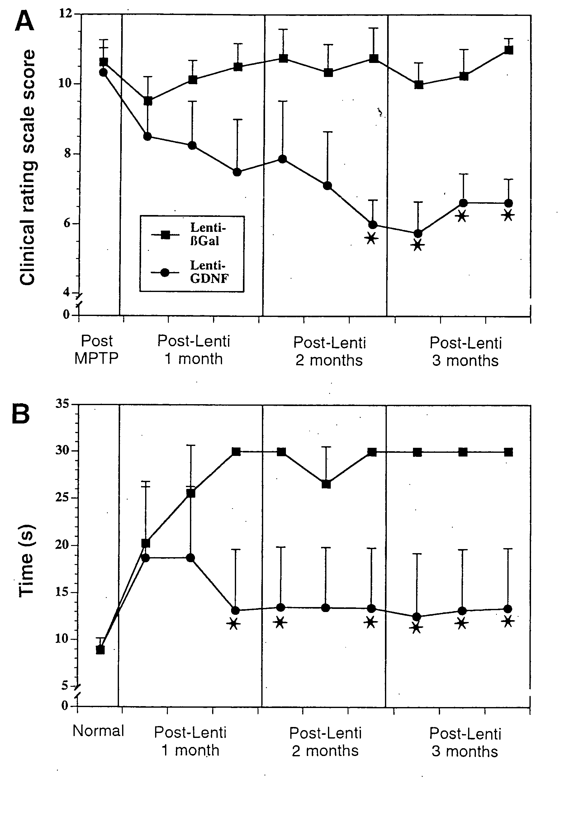
[0055] Non-lesioned aged monkeys that model PD like neurodegeneration (17) display a slow progressive loss of dopamine within the striatum and tyrosine hydroxylase (TH) within the substantia nigra without frank cellular degeneration. Eight aged (approximately 25 years old) female rhesus monkeys received injections of lentiviral vectors encoding—galactosidase (lenti-Gal; n=4) or GDNF (lenti-GDNF; n=4) targeted for the striatum and substantia nigra and were killed 3 months later.

[0056] Under MRI guidance, each monkey received six stereotaxic injections of lenti-Gal or lenti-GDNF bilaterally into the caudate nucleus, putamen, and substantia nigra. Injections were made into the head of the caudate nucleus (10 μl), body of the caudate nucleus (5 μl), anterior putamen (10 μl), commissural putamen (10 μl), postcommissural putamen (5 μl), and substantia nigra (5 μl). Injections were made...

example iii

DOPA Uptake Following GDNF Treatment in Aged Animals

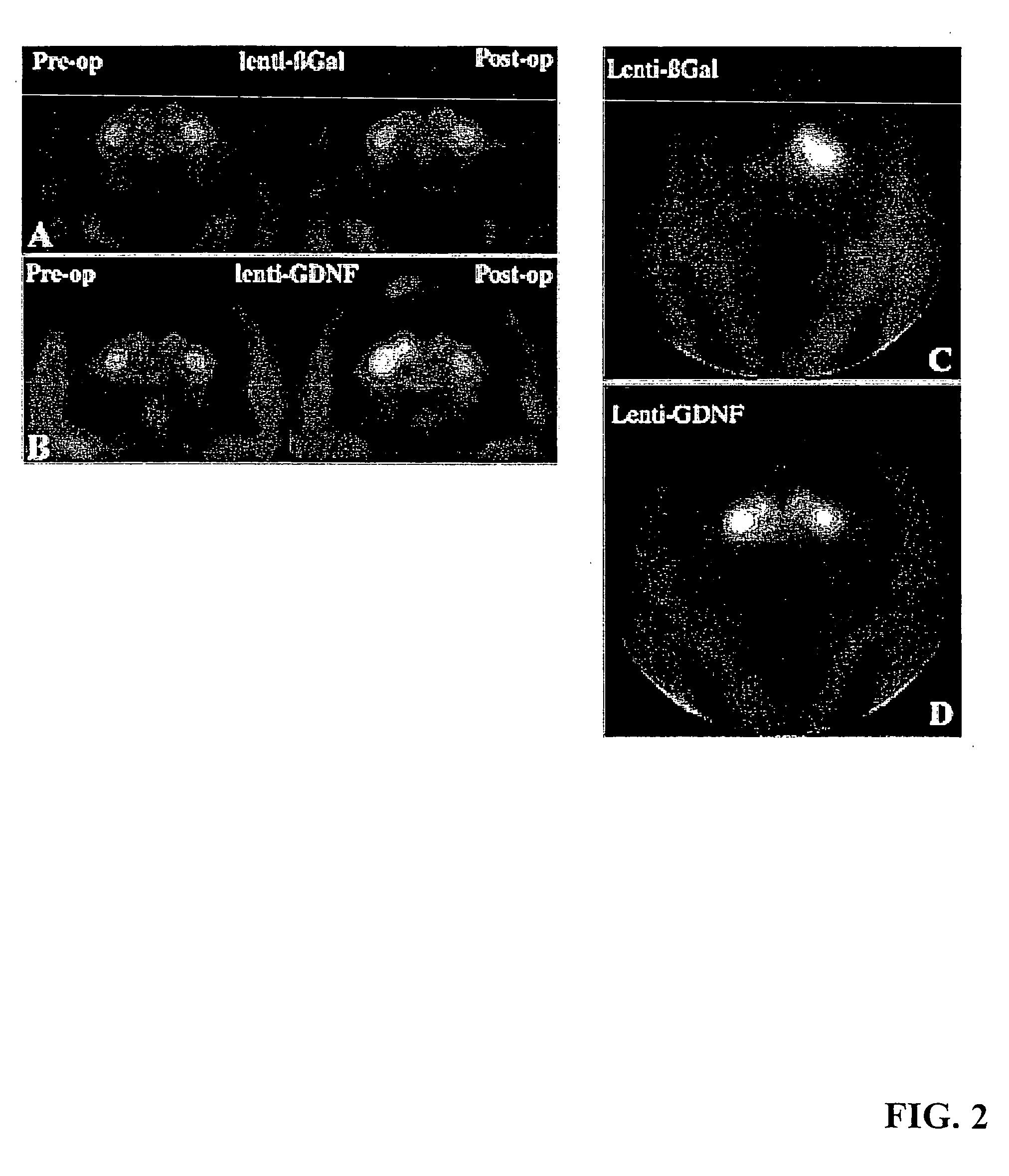
[0061] Aged monkeys underwent fluorodopa (FD) positron emissiontomography (PET) before surgery and again just before being killed. Before treatment, all monkeys displayed symmetrical FD uptake in the caudate and putamen bilaterally (ratio: 1.02±0.02) (FIGS. 2A and 2B, left). Similarly, there was symmetrical (4% difference) FD uptake in all lenti-Gal-treated monkeys after lentivirus injections (FIG. 2A, right).

[0062] In contrast, FD uptake was significantly asymmetrical (27%) in lenti-GDN-F-treated monkeys with greater uptake on the side of the GDNF expression (P<0.007; FIG. 2B, right). With respect to absolute values, lenti-Gal animals displayed a trend toward reduced FD uptake after treatment relative to baseline levels (P=0.06). Qualitatively, three of four lenti-GDNF-treated monkeys displayed clear increases in FD uptake on the treated side.

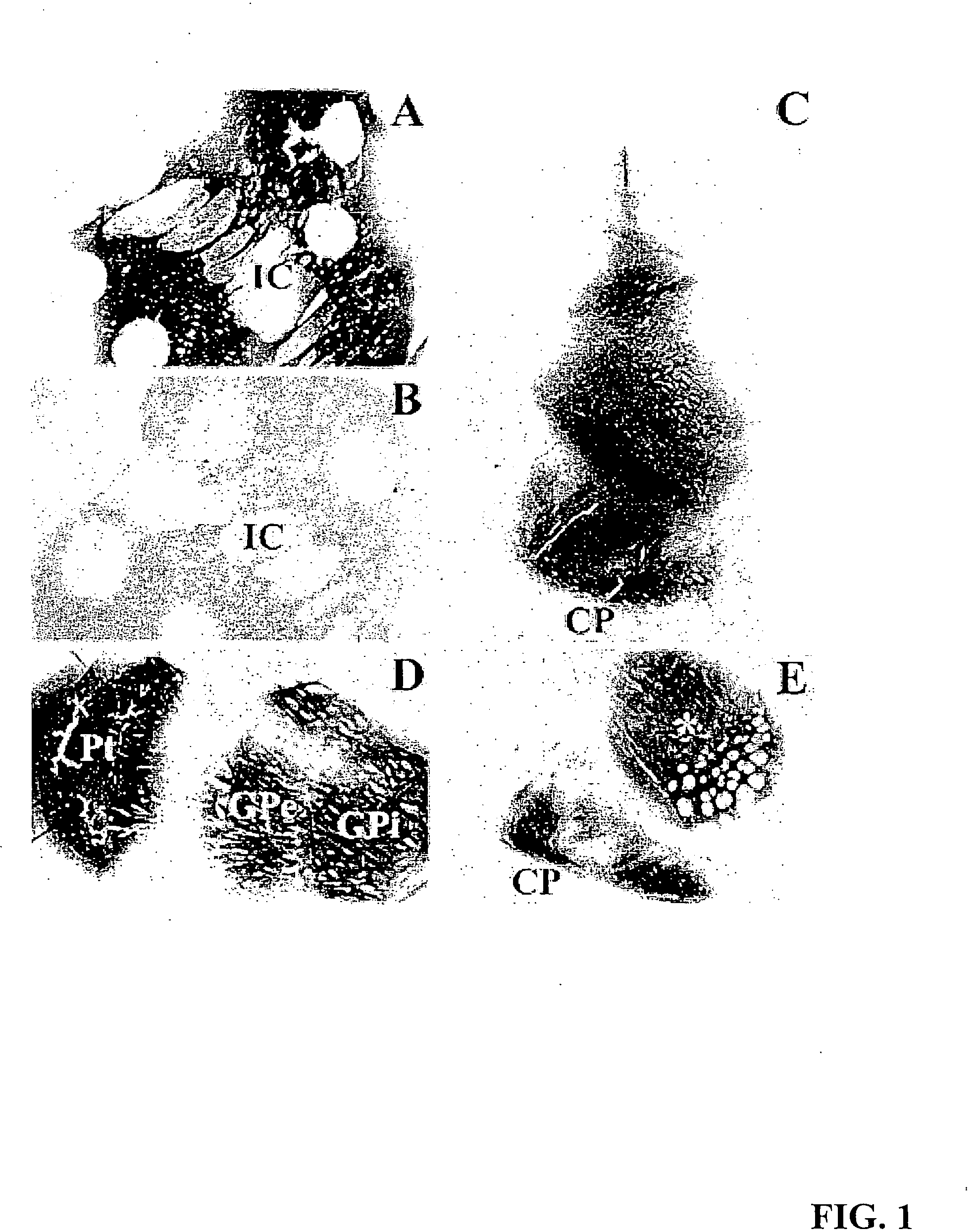
[0063] Within the striatum, lentiviral delivery of GDNTF increased a number of markers...


PUM


Abstract

A specific clinical protocol for use toward therapy of defective, diseased and damaged neurons in the mammalian brain, of particular usefulness for treatment of neurodegenerative conditions such as Parkinson's disease and Alzheimer's disease. The protocol is practiced by delivering a definite concentration of recombinant neurotrophin, such as glial cell-derived neurotrophic factor), into a targeted region of the brain (such as the substantia nigra) using a lentiviral expression vector. The neurotrophin is delivered to, or within close proximity of, identified defective, diseased or damaged brain cells. The concentration of neurotrophin delivered as part of a neurotrophic composition varies from 1010 to 1015 neurotrophin encoding viral particles / ml of composition fluid. Each delivery site receives from 2.5 μl to 25 μl of neurotrophic composition, delivered slowly, as in over a period of time ranging upwards of 10 minutes / delivery site. Each delivery site is at, or within 500 μm of, a targeted cell, and no more than about 10 mm from another delivery site. The method stimulates growth of targeted neurons, and reversal of functional deficits associated with the neurodegenerative disease being treated.

Description

RELATED U.S. PATENT APPLICATIONS [0001] This is a continuation of, and claims priority of, U.S. patent application Ser. No. 10 / 032,952, filed on Oct. 26, 2001, now U.S. Pat. No. 6,815,431, issued on Nov. 9, 2004, which is a continuation-in-part of, and claims the priority of U.S. patent application Ser. No. 09 / 620,174, filed on Jul. 19, 2000, now U.S. Pat. No. 6,683,058, issued on Jan. 27, 2004, which is a continuation-in-part of, and claims the priority of U.S. patent application Ser. No. 09 / 060,543, filed on Apr. 15, 1998, now U.S. Pat. No. 6,451,306.FIELD OF THE INVENTION [0002] The invention relates to methods for treatment of neurodegenerative disease and methods for delivery of therapeutic neurotrophins into the mammalian brain. HISTORY OF THE RELATED ART [0003] Neurotrophins play a physiological role in the development and regulation of neurons in mammals. In adults, basal forebrain cholinergic neurons, motor neurons and sensory neurons of the CNS retain responsiveness to neu...

Claims


Application Information

Patent Timeline
Patent Type & Authority Applications(United States)
IPC IPC(8): A61K38/18A61K48/00C07K14/475
CPCA61K38/185A61K48/00A61K48/0075A61K48/0083C07K14/475A61K48/005C12N2740/16043C12N2799/021C12N15/867C12N2799/027C12N15/86
Inventor TUSZYNSKI, MARK H.
Owner RGT UNIV OF CALIFORNIA
Why Eureka
Try Eureka