Method for radiological image processing

a radiological image and processing technology, applied in the field of mammography, can solve problems such as eye fatigue, affecting the ability to perceive clusters, and inconvenient use of these systems, and affecting the ability of radiologists to replace radiologists

Inactive Publication Date: 2005-06-23
GE MEDICAL SYST GLOBAL TECH CO LLC
View PDF26 Cites 22 Cited by
  • Summary
  • Abstract
  • Description
  • Claims
  • Application Information

AI Technical Summary

Benefits of technology

[0006] An embodiment of the method of the invention allows enhancing all the signs of micro-calcification that appear on the image without distinction, whether these signs are grouped or isolated, benign or malignant. Consequently, the radiologist obtains an immediate overall view of the distribution mapping of micro-calcifications. The radiologist can then directly observe the shape and extension of clusters without necessarily needing to zoom to the areas in question.
[0008] In an embodiment of the invention, the enhanced intensity of elements that may constitute micro-calcifications is performed on a “presentation” image that can be viewed by the radiologist and is distinct from the raw image from which the elements are detected. This presentation image can be obtained through other processing methods designed to improve the ease with which the radiologist reads the image.

Problems solved by technology

Nevertheless, some radiologists do not perceive certain deposits.
Furthermore, radiologists must often analyze several mammography images one after the other, which could lead to eye fatigue that could affect their capacity to perceive clusters.
The inconvenience of these systems is that they tend to replace the radiologist in the interpretation of the image, in particular, in terms of distinguishing between malignant elements and benign elements.
Nevertheless, these systems are not sufficiently reliable to completely replace radiologists.

Method used


Image

  • Method for radiological image processing
  • Method for radiological image processing
  • Method for radiological image processing

Examples


Embodiment Construction

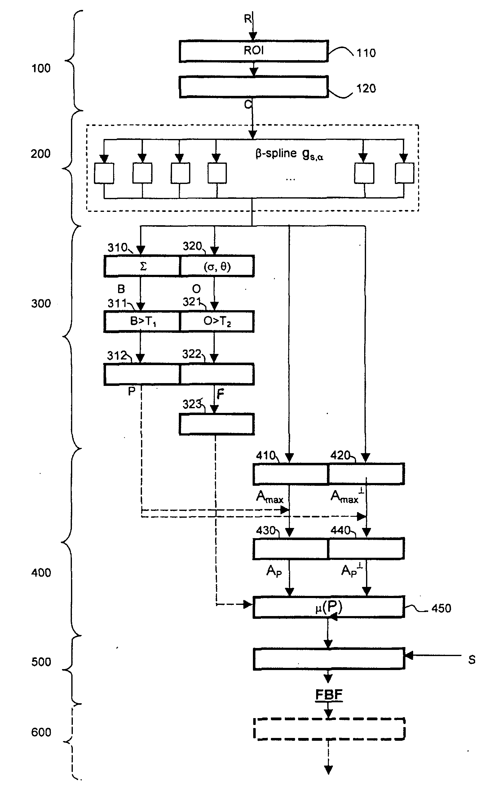
[0014] An embodiment of the invention is represented in FIG. 1 where the image processing method is applied to a raw image R; that is, it is provided directly by a digital detector of a radiology system without having been pre-processed. It is from this raw image R that the processing method allows locating elements or objects that may constitute signs of micro-calcification.

[0015] The processing method also uses an image called a “presentation image” on which the elements will be enhanced in the end. This presentation image S is obtained by another processing method from the raw image R. This method allows providing an image that can be viewed directly by the radiologist. EP 1 113 392 describes, for example, a method for thickness compensation that allows adapting the grey scale variations for displaying an image whose appearance allows performing a reading along the entire extension of the patient's breast.

[0016] In FIG. 1, the image processing method may include five phases: (1...


PUM


Abstract

A method for radiological image processing that includes: locating, on a radiological image provided by an imaging device, elements that may constitute signs of micro-calcification and displaying an image on which the intensity of elements thus localized is enhanced.

Description

CROSS-REFERENCE TO RELATED APPLICATIONS [0001] This application claims the benefit of a priority under 35 USC 119(a)-(d) to French Patent Application No. 03 14457 filed Dec. 10, 2003, the entire contents of which are hereby incorporated by reference. BACKGROUND OF THE INVENTION [0002] This invention relates to the field of radiological imaging and, more specifically, an object of interest particularly in the field of mammography. Mammography is largely used for detecting lesions and preventing breast cancer. The first signs that radiologists search for on mammogram images are calcium deposits called micro-calcifications, which generally constitute more opaque elements in X-rays than the surrounding tissue. Radiologists scrutinize the images looking for signs of these micro-calcifications. Radiologists can, to a certain degree, differentiate between malignant micro-calcifications (often a group of clusters) and benign calcium deposits. Radiologists interpret the shape and brightness ...

Claims


Application Information

Patent Timeline
Patent Type & Authority Applications(United States)
IPC IPC(8): G06T1/00G06T5/00A61B6/00G06T5/10G06T5/20G06T7/00G06T7/40G06V10/25H04N5/325
CPCG06K9/3233G06K2209/053G06T5/10G06T2207/20012G06T7/0012G06T2207/30068G06T5/008G06T5/20G06V10/25G06V2201/032
Inventor BERNARD, SYLVAINMULLER, SERGEBOUCHEVREAU, XAVIER
Owner GE MEDICAL SYST GLOBAL TECH CO LLC
Why Eureka
Try Eureka