Method and system of treatment of heart failure using 4D imaging

a heart failure and imaging technology, applied in the field of heart failure treatment methods and systems, can solve the problems of significant cost, poor quality of life of patients with chf, and a major health problem worldwid

Inactive Publication Date: 2005-06-30
MEDTRONIC INC
View PDF99 Cites 67 Cited by
  • Summary
  • Abstract
  • Description
  • Claims
  • Application Information

AI Technical Summary

Problems solved by technology

Despite considerable progress in the management of congestive heart failure (CHF), it remains a major health problem worldwide.
Despite significant advances in the treatment of CHF using various pharmacological therapies, quality-of-life in patients with CHF is poor as they are frequently hospitalized and heart failure is a common cause of death.
In addition, there is significant cost attached to this problem.
There are, however, several problems with this approach.
Initially, this type of approach is time-consuming for the physician.
Placement of the left ventricular lead is limited to sites available that provide reasonable pacing and sensing parameters.
Cannulating the coronary sinus can be challenging due to enlarged right atrium, rotation of the heart and presence of Tebesian valve (a valve close to the opening of the coronary sinus).
In most instances, problems with the placement of the coronary sinus lead are identified at the time of the interventional procedure.
Unfortunately, there are many problems with epicardial lead placement as well, some of which include but are not limited to: I) Limited view of the posterola

Method used


Image

  • Method and system of treatment of heart failure using 4D imaging
  • Method and system of treatment of heart failure using 4D imaging
  • Method and system of treatment of heart failure using 4D imaging

Examples


Embodiment Construction

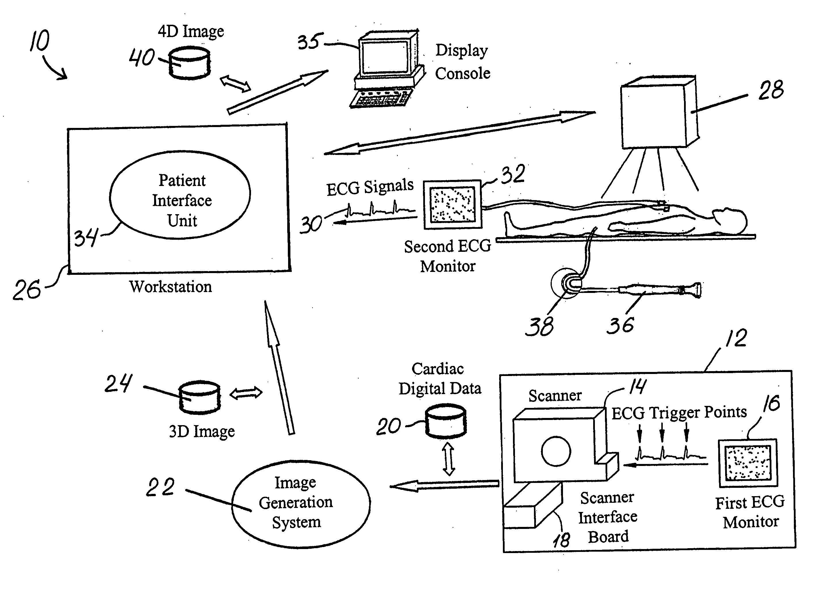
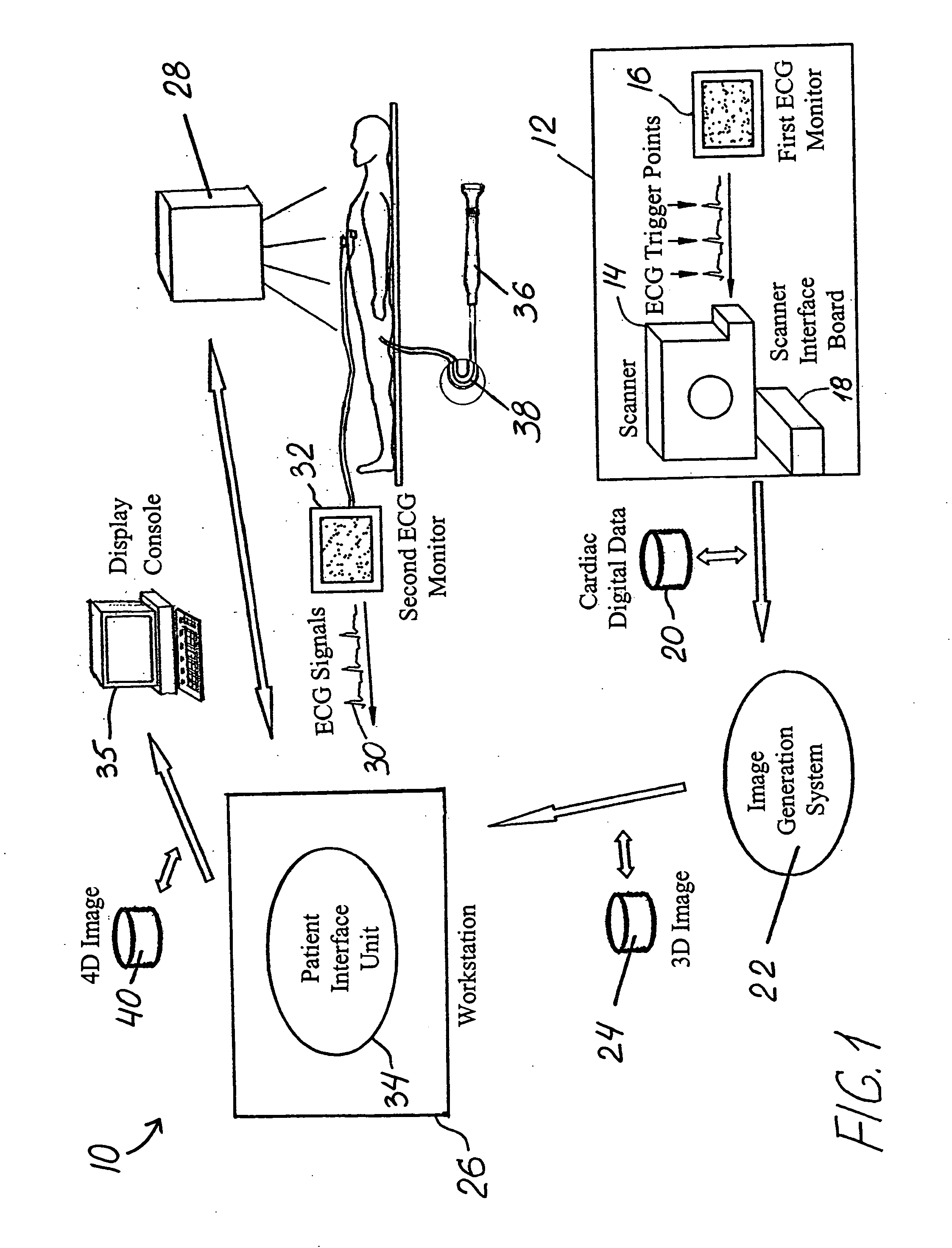
[0032] The drawings illustrate embodiments of a system and method for treating heart failure in a patient using 4D imaging in accordance with this invention. The embodiments shown enable an electrophysiologist, cardiologist and / or surgeon to plan in advance and to later perform an interventional procedure such as bi-ventricular pacing in a manner that makes the procedure simpler and more efficacious while decreasing the risk of complications.

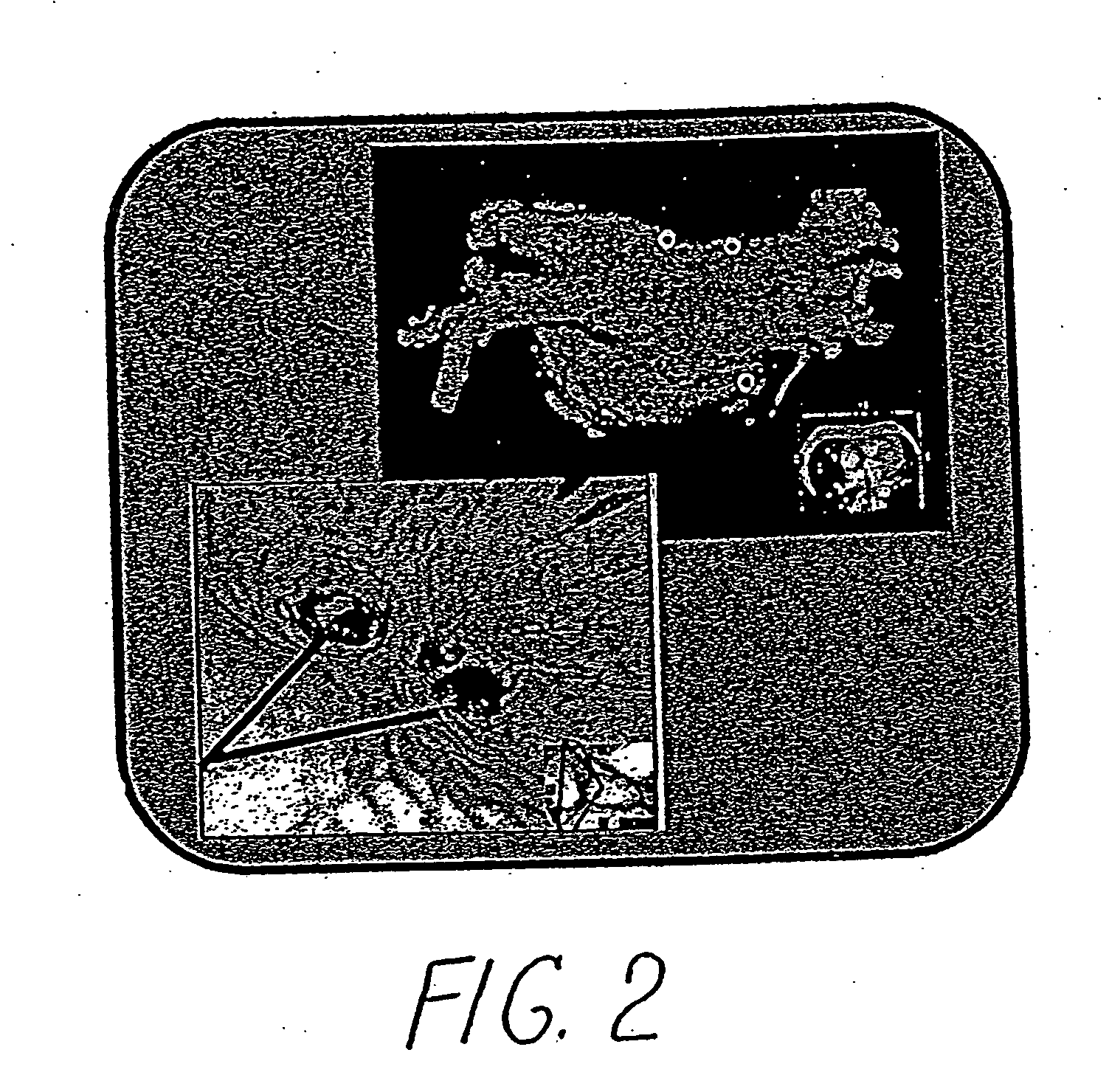
[0033] Using imaging systems known in the art, 3D images are obtained of a cardiac chamber such as the left ventricle and the adjacent coronary sinus. These images include detailed 3D models of the left ventricle and endocardial views (i.e., navigator or views from the inside) of the coronary sinus. These images are then registered and synchronized with real-time cardiac motion on an interventional system such as a fluoroscopic system to generate a 4D image. In this manner, detailed 3D images acquired at different phases of the cardiac cycle pr...


PUM


Abstract

A method is provided for treatment of heart failure having the steps of obtaining cardiac digital data from a medical imaging system utilizing an ECG gated protocol; generating a series of 3D images of a cardiac chamber and its surrounding structures, preferably the left ventricle and coronary sinus, from this cardiac digital data at select ECG trigger points that correspond to different phases of the cardiac cycle; registering these 3D images with an interventional system; acquiring ECG signals from the patient in real-time; transmitting these ECG signals to the interventional system; synchronizing the registered 3D images with trigger points on the transmitted ECG signals to generate a 4D image; visualizing this 4D image upon the interventional system in real-time; visualizing a pacing/defibrillation lead over the 4D image upon the interventional system; navigating the pacing/defibrillation lead utilizing the 4D image; and placing the pacing/defibrillation lead over the cardiac chamber at an appropriate site to treat the heart failure.

Description

RELATED APPLICATION [0001] This application claims the benefit of U.S. Provisional Applications Nos. 60 / 531,296, 60 / 531,293 and 60 / 531,294, each filed on Dec. 19, 2003 and the contents of each are incorporated by reference herein in its entirety.FIELD OF THE INVENTION [0002] This invention relates generally to methods and systems for treatment of heart failure using bi-ventricular pacing / defibrillation leads and, in particular, to methods and systems utilizing 3D digital images for cardiac interventional procedures in such treatment and for the planning of such procedures. BACKGROUND OF INVENTION [0003] Despite considerable progress in the management of congestive heart failure (CHF), it remains a major health problem worldwide. It is estimated that there are 6-7 million people with CHF in the United States and Europe and approximately 1 million patients are diagnosed with CHF every year. [0004] Despite significant advances in the treatment of CHF using various pharmacological thera...

Claims


Application Information

Patent Timeline
IPC IPC(8): A61B17/00A61B19/00G06T7/00G06T17/00G16H20/30G16H30/40
CPCA61B5/7285A61B6/506A61B8/543A61B19/52A61B2017/00039A61B2017/00703A61B2019/5238A61N1/3627G06F19/3406G06F19/3437G06F19/3481G06T7/0012G06T7/0038G06T17/00G06T2207/10076G06T2207/10121G06T2207/30048A61B6/541A61B90/36A61B2090/376G06T7/38G16H40/63G16H50/50G16H30/40G16H20/30
Inventor SRA, JASBIR S.
Owner MEDTRONIC INC
Why Eureka
Try Eureka