

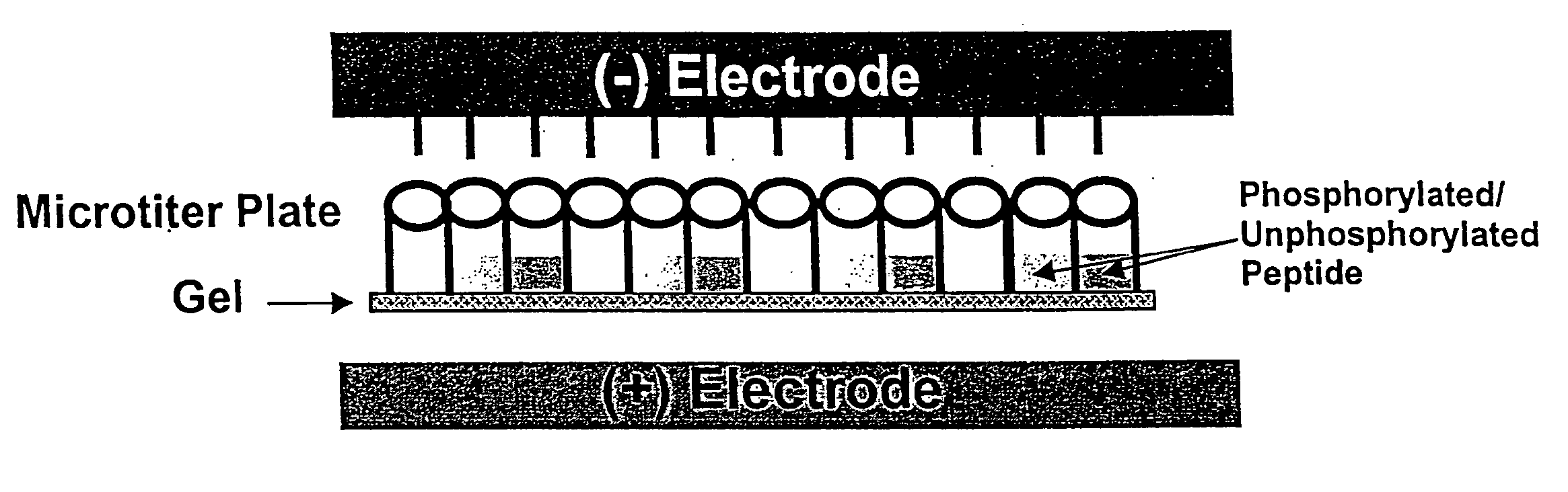
Microtiter plate format device and methods for separating differently charged molecules using an electric field
a microtiter plate and electric field technology, applied in the direction of fluid pressure measurement, liquid/fluent solid measurement, peptides, etc., can solve the problems of high quantity of sup>32/sup>p to be used in assays, significant disadvantages, and waste cannot be easily disposed of, so as to achieve the effect of convenient adaptation
- Summary
- Abstract
- Description
- Claims
- Application Information
AI Technical Summary
Benefits of technology
Problems solved by technology
Method used
Image

Examples
example 1
Illustrative Assay for Protein Kinase A Phosphorylation of Substrate Peptide in an Azarose- or Acrylamide-Filled Electrophoresis Sample Plate
[0051] Reagents: [0052] 20 mM Tris-HCl pH 8.0 [0053] 10 mM MgCl2 [0054] 1 mM ATP [0055] 1 μM cAMP [0056] 60 μM Kemptide [0057] 350 mM K3PO4 pH 7.5 [0058] 0.1 mM DTT [0059] 0.8% agarose gels in 50 mM Tris-HCl, pH 8.0 or [0060] 8%, and 20% acrylamide gels (19:1 Acrylamide:Bis-acrylamide), with 0.5% Darocure 4265
[0061] Agarose- or acrylamide-filled electrophoresis sample plates were prepared by the following methods: Sample plates of microtiter wells open on both ends were sealed on the bottom end with a Dynex Technologies plate sealer. 0.8% agarose in 50 nM Tris-Cl pH 8.0 was melted to a fluid consistency. While hot, the agarose was pipetted into the bottom of each well of the sealed sample plate. 96-well sample plates were filled with 100 μl agarose and 384-well sample plates were filled with 301 μl per well. After about 20 minutes, when the a...
example 2
Illustrative Assay For Protein Kinase A Phosphorylation of Substrate Peptide in a Gel / Membrane Sample Plate with Conductive Liquid Electrodes
[0065] Acrylamide / membrane sample plates were prepared from 384-well plates (produced by Greiner) with Biodyne B membrane (from Pall, Inc.) on the bottom of the wells. 15 μl of 20% acrylamide was pipetted into each well and UV cured, as described above, to form a diffusion-inhibiting layer.
[0066] Samples of phosphorylated and unphosphorylated Texas Red labeled Kemptide were then prepared as described above in Example 1. Samples of charged peptides (20 μl of 10 μM, or 50 mmol peptide) were diluted into 1× Tris-borate buffer (pH8.0) and applied into the wells of the 384 well sample plate.
[0067] Conductive-liquid second electrodes, as shown in FIG. 4, were used. The top electrode reservoir was filled with the Tris-borate buffer or 50 mM Tris-HCl. Electrophoresis was carried out for 5 minutes at 100 mAmp. The wells were then washed and read usin...
example 3
[0069] Electrophoresis in 1536-well Sample Plates Using Gel-Capillary Electrodes Reaction plates for this device were made by Greiner with a Biodyne B membrane coating the bottom of the plate. A 3 μl layer of 20% acrylamide served as a diffusion-inhibiting layer at the bottom of each well. A gel-capillary upper structure was designed to allow current to pass through to the wells while segregating the electrochemistry at the cathode from the reaction mixture, as shown in FIG. 6.
[0070] For effective electrophoresis, the microcapillaries were filled with a gel that has low resistance and also prevents the buffer bath from leaking into the reaction plate. We tested agarose gels with concentrations ranging from 0.3 to 1.0%. These gels had low resistance, with currents of 0.3 to 0.6 mAmps / well. To make more robust gels, agarose was chemically crosslinked to the interior surface of the glass microcapillary. A solution of 1% N-(2-aminoethyl)-3-aminopropyl-trimethoxysilane (AEAPS) was prepa...
PUM
| Property | Measurement | Unit |
|---|---|---|
| Length | aaaaa | aaaaa |
| Length | aaaaa | aaaaa |
| Fraction | aaaaa | aaaaa |
Abstract
Description
Claims
Application Information