
Method and system for intelligent qualitative and quantitative analysis of digital radiography softcopy reading

a digital radiography and softcopy reading technology, applied in the field of diagnostic information analysis, can solve the problems of difficult observation of objects, inability to provide quantitative measurements, and complex radiographic images

Active Publication Date: 2005-09-08
EDDA TECH
View PDF17 Cites 38 Cited by
  • Summary
  • Abstract
  • Description
  • Claims
  • Application Information

AI Technical Summary

Benefits of technology

The patent is about a system and method for assisting physicians in reading digital / digitized radiographic images. The current systems available are not capable of providing quantitative measurements, complying with a clinical workflow, or offering assistance based on physician-specific knowledge and experience. The invention aims to address these issues by providing a system that can provide more accurate and reliable quantitative measurements, better comply with a clinical workflow, and offer assistance based on physician-specific knowledge and experience. The invention uses a combination of machine learning and artificial intelligence techniques to analyze and segment radiographic images, detect and locate nodules, and provide assistance to physicians in making diagnostic decisions. The invention also includes a user-friendly interface that allows for real-time interaction and collaboration with the system.

Problems solved by technology

Most radiographic images are complex due to the fact that three dimensional anatomical structures are projected on a two dimensional image plane.
These may cause difficulty to observe the object(s) of interest and discern the boundary of such object(s).
First, most of the existing systems are not capable of providing quantitative measurements, which are often used by physicians to reach a diagnostic decision.
This incapability is often related to the difficulties in segmenting out nodules and / or lesions in images when structural / anatomic noise exists due to, for example, the difficulties stated above.
Second, existing systems are not capable of complying with an existing clinical workflow and provide only assistance in certain stages of such a workflow.
Third, existing systems usually employ black-box approaches so that it is not possible for physicians to interact in real time with such systems.
As a consequence, such systems can provide only assistance based on prior knowledge that is built in the system rather than offering assistance based on physician-specific knowledge and experience.

Method used


Image

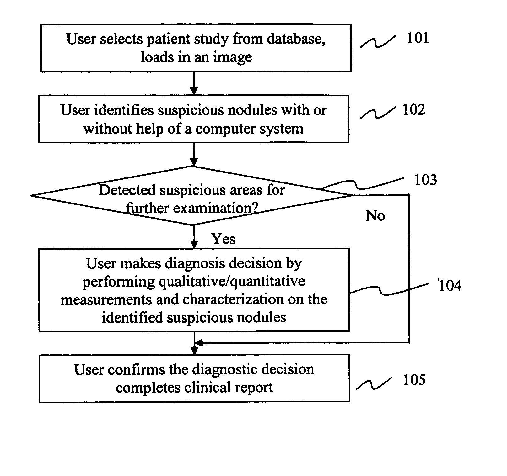
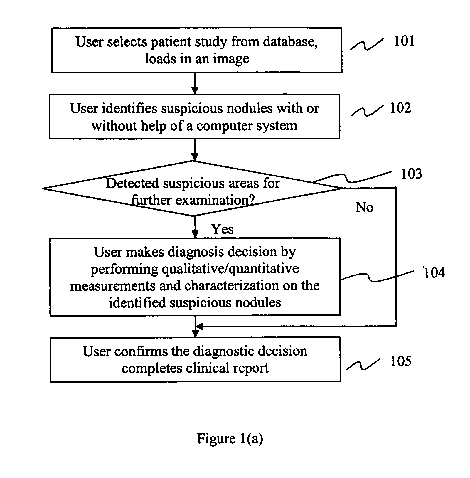
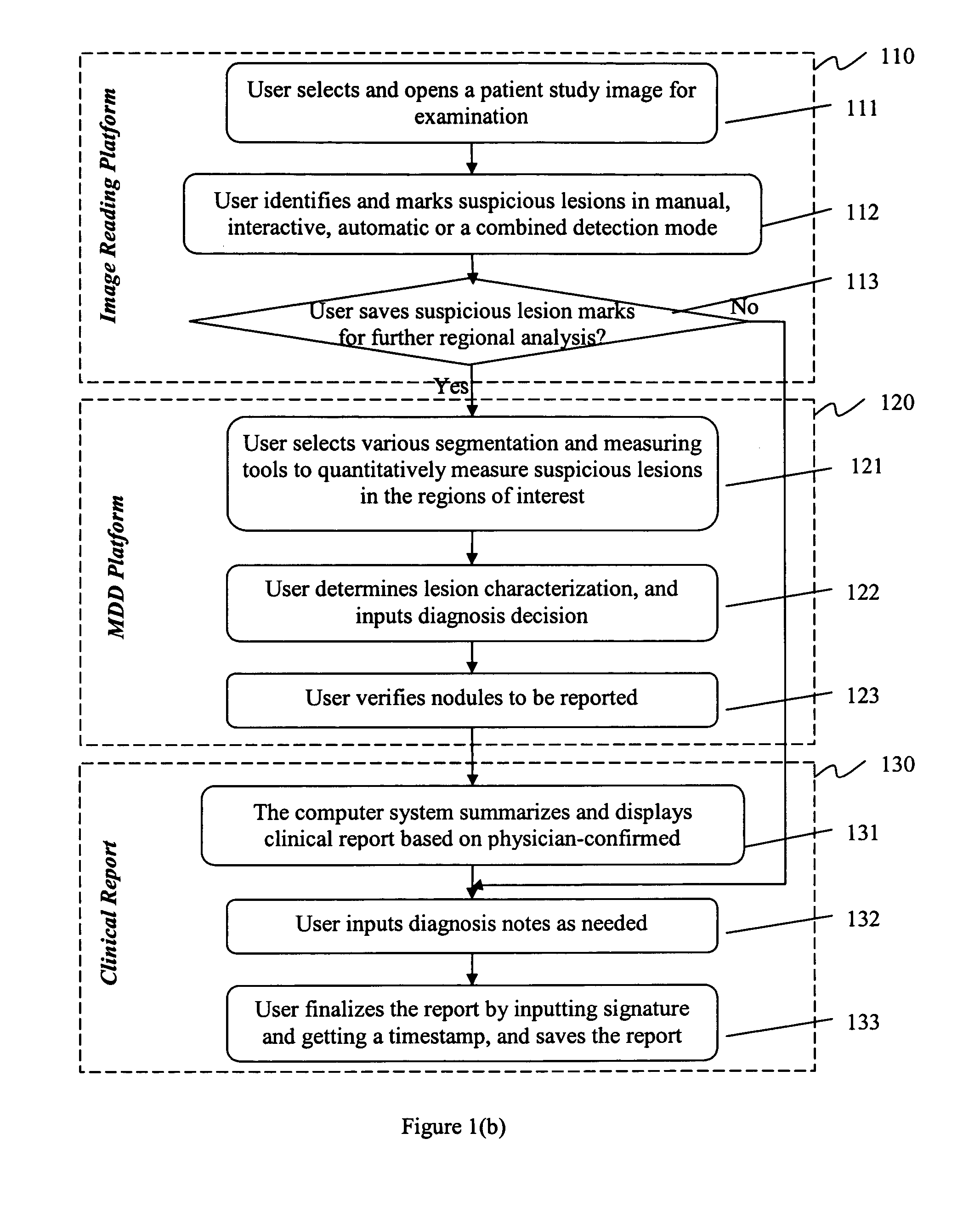
  • Method and system for intelligent qualitative and quantitative analysis of digital radiography softcopy reading
  • Method and system for intelligent qualitative and quantitative analysis of digital radiography softcopy reading
  • Method and system for intelligent qualitative and quantitative analysis of digital radiography softcopy reading

Examples


Embodiment Construction

[0041] The processing described below may be performed by a properly programmed general-purpose computer alone or in connection with a special purpose computer. Such processing may be performed by a single platform or by a distributed processing platform. In addition, such processing and functionality can be implemented in the form of special purpose hardware or in the form of software or firmware being run by a general-purpose or network processor. Data handled in such processing or created as a result of such processing can be stored in any memory as is conventional in the art. By way of example, such data may be stored in a temporary memory, such as in the RAM of a given computer system or subsystem. In addition, or in the alternative, such data may be stored in longer-term storage devices, for example, magnetic disks, rewritable optical disks, and so on. For purposes of the disclosure herein, computer-readable media may comprise any form of data storage mechanism, including such...


PUM


Abstract

The present invention describes a method and system for intelligent diagnostic relevant information processing and analysis. Information associated with a patient is processed via an image reading platform. Based on such processed information, a matrix of diagnosis decisions containing diagnostic related information is generated via a matrix of diagnosis decision platform. A diagnostic decision is made based on the diagnostic relevant information. The image reading platform and / or the matrix of diagnosis decision platform encapsulate information and toolkits to be used to manipulate the information.

Description

CROSS-REFERENCE TO RELATED APPLICATIONS [0001] The current Application is based on U.S. Provisional Application No. 60 / 537,558 filed Jan. 21, 2004, and U.S. Provisional Application No. 60 / 562,260 filed Apr. 15, 2004, the entire contents of which applications are hereby incorporated by reference.BACKGROUND OF THE INVENTION [0002] 1. Field of the Invention [0003] The invention disclosed herein relates to a system and method for assisted medicine. Particularly, it relates to diagnostic information analysis. [0004] 2. Description of Related Art [0005] Most radiographic images are complex due to the fact that three dimensional anatomical structures are projected on a two dimensional image plane. For example on chest radiographic images, over 60 percent of the lung region may be occluded by ribs. Object(s) of interest such as nodules may therefore overlap with anatomical structures such as ribs, reside in shadows, or may be occluded by other types of objects. These may cause difficulty to...

Claims


Application Information

Patent Timeline
Patent Type & Authority Applications(United States)
IPC IPC(8): A61B5/05G06T7/00G16H15/00G16H30/20G16H30/40G16H50/20
CPCG06T7/0012G06T2200/24G06T2207/10116G06T2207/30061A61B6/50G06T7/0079G06F19/321G06F19/3406G06F19/3487G06T5/008A61B6/5217G06T7/10G16H40/63G16H15/00G16H30/40G16H50/20G16H30/20G06T5/94
Inventor QIAN, JIANZHONGFAN, LIWEI, GUO-QINGLIANG, CHENG-CHUNG
Owner EDDA TECH
Learn More