Neoplasm specific antibodies and uses thereof
a technology of neoplasm and specific antibodies, applied in the field of cancer diagnosis and treatment, can solve the problems of poor prognosis in 50 years, and achieve the effect of inhibiting the proliferation of neoplastic cells
- Summary
- Abstract
- Description
- Claims
- Application Information
AI Technical Summary
Benefits of technology
Problems solved by technology
Method used
Image
Examples
example 1
Materials and Methods
Producing Hybridomas
[0095] We immortalized lymphocytes by fusing them to the HAB-1 heteromyeloma as follows.
[0096] We washed the HAB- 1 heteromyeloma cells twice with RPMI 1640 (PAA, Vienna, Austria) without additives and centrifuged the cells for 5 minutes at 1500 rpm. We then thawed frozen lymphocytes obtained from either the spleen or the lymph nodes and we washed these cells twice with RPMI 1640 without additives and centrifuged these cells at 1500 rpm for 5 minutes. Both the HAB-1 and the lymphocyte cell pellets were resuspended in 10 ml RPMI 1640 without additives and were counted in a Neubauer cell counting chamber. We washed the cells again, added the HAB-1 cells and the lymphocytes together in a ratio of 1:2 to 1:3, mixed them, and centrifuged the mixture for 8 minutes at 1500 rpm. We pre-warmed Polyethylene Glycol 1500 (PEG) to 37° C. and carefully let the PEG run drop-wise onto the pellet while slightly rotating the 50 ml tube. Next, we gently res...
example 2
Generation of the Cell Line Expressing the CM-1 Monoclonal Antibody
[0114] As described above, we obtained the CM-1 monoclonal antibody expressing hybridoma by fusing lymphocytes obtained from the spleen of a patient having a moderately differentiated colorectal adenocarcinoma (tumor staging T2N0Mx, grade 2) with the heteromyeloma cell line HAB-1 (Faller, et a., Br. J. Cancer 62:595-598, 1990). The resultant cell is a type of hybridoma known as a trioma, as it is the fusion of three cells. Like normal B-lymphocytes, this trioma has to ability to produce antibodies. The specificity of the antibody is determined by the specificity of the original lymphocyte from the patient that was used to generate the trioma.
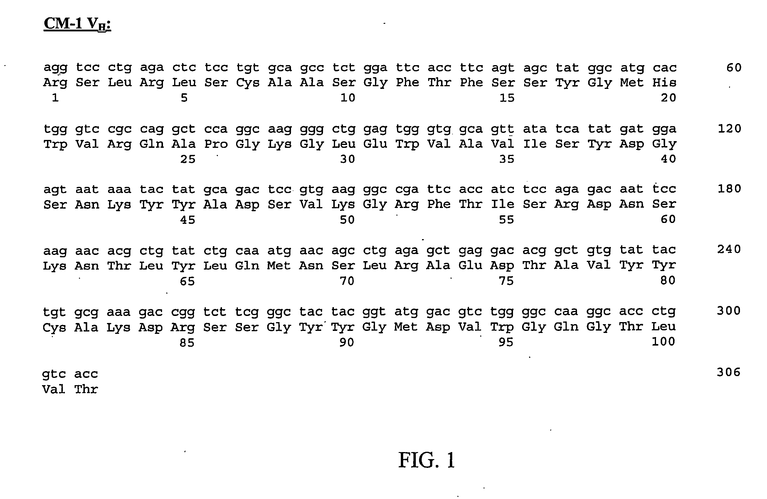
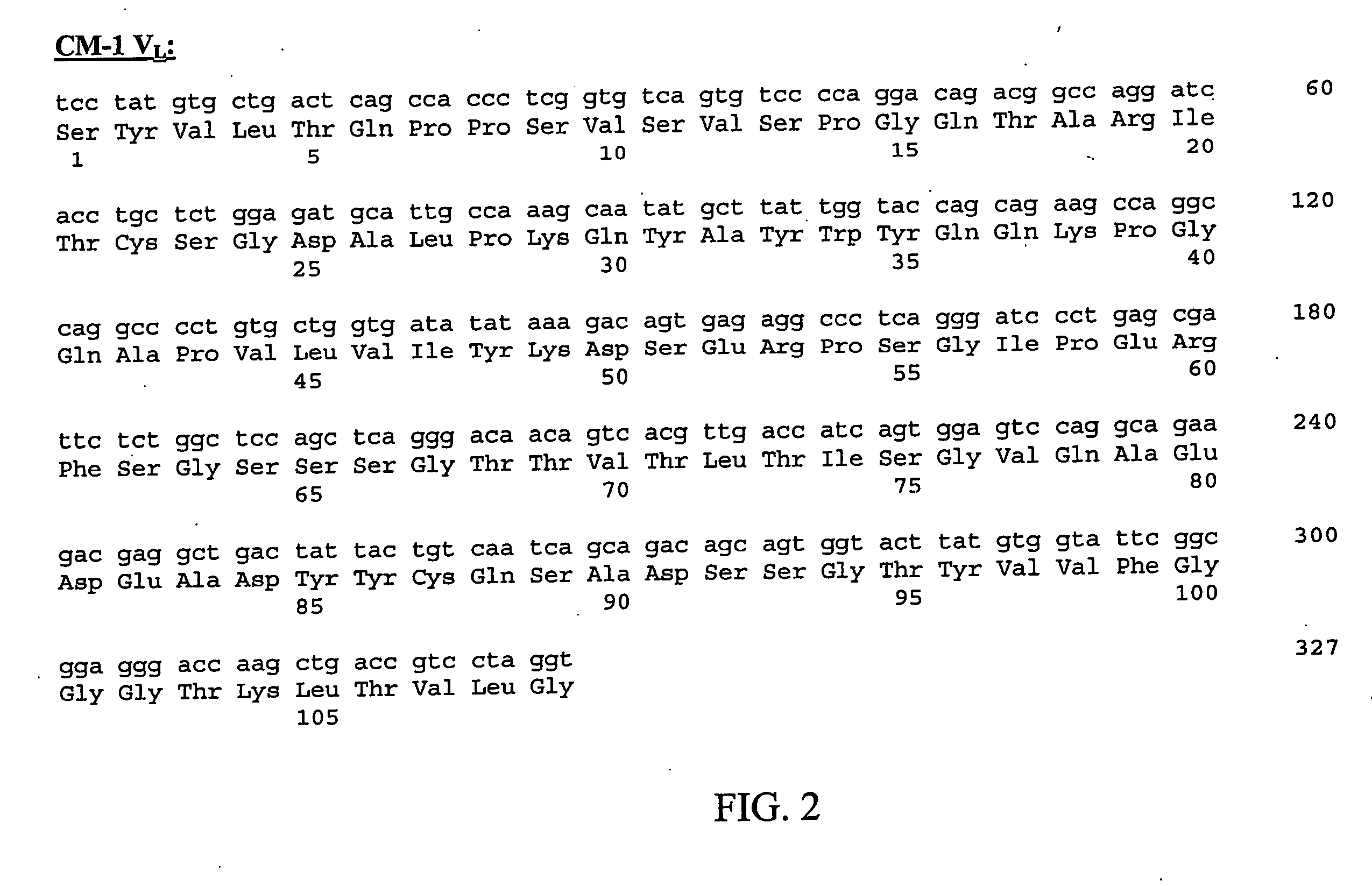
[0115] The amino acid and nucleic acid sequences of the CM-1 monoclonal antibody VH region are shown in FIG. 1 as SEQ ID NO:1 and SEQ ID NO:2, respectively, and the amino acid and nucleic acid sequences of the CM-1 VL region are shown in FIG. 2 as SEQ ID NO:3 and SEQ ID NO:4, r...
example 3
Immunohistochemical Characterization of an Antibody
[0116] To characterize the monoclonal antibody secreted by a hybridoma, we tested the antibody against a panel of normal and tumor tissues using an immunoperoxidase assay as described in the materials and methods. This assay provided us with an overview of which tissues were stained by the antibody and of the distribution of the antigen.
[0117] Antibodies that are specific for tumor cells and not for normal tissue were further characterized. First, we tested these antibodies against the same types of tumors from different patients. We then tested these antibodies against tumors of other organs and, finally, against normal tissues. Using these assays, we identified the human CM-1 monoclonal antibody.
[0118] The CM-1 monoclonal antibody specifically stains neoplastic cells, such as colorectal carcinoma cells. We stained tissue samples obtained from 21 different colorectal tumors with the CM-1 monoclonal antibody. 10 of these tumors (...
PUM
| Property | Measurement | Unit |
|---|---|---|
| Cell proliferation rate | aaaaa | aaaaa |
Abstract
Description
Claims
Application Information
 - R&D
- Intellectual Property
- Life Sciences
- Materials
- Tech Scout
- Unparalleled Data Quality
- Higher Quality Content
- 60% Fewer Hallucinations
Browse by: Latest US Patents, China's latest patents, Technical Efficacy Thesaurus, Application Domain, Technology Topic, Popular Technical Reports.
© 2025 PatSnap. All rights reserved.Legal|Privacy policy|Modern Slavery Act Transparency Statement|Sitemap|About US| Contact US: help@patsnap.com


