Endoscopic fastening system with multiple fasteners

a technology of multiple fasteners and endoscopic fasteners, which is applied in the field of endoscopic fastening devices and surgical fasteners, can solve the problems of no reliable method for securing fasteners inside the patient's body, no such devices currently available, and the limitations of suturing operations are the sam

Inactive Publication Date: 2005-09-29
GRANIT MEDICAL INNOVATION
View PDF5 Cites 293 Cited by
  • Summary
  • Abstract
  • Description
  • Claims
  • Application Information

AI Technical Summary

Benefits of technology

[0029] The above-described method is an important advance in the technology of small fasteners (staples, clips, etc) because it precludes the need for the “opened-closed-opened” technique. This method, described herein for the first time, enables the “closed-closed-closed” technique, thereby simplifying the design of an endoscopic fastening system to the extent of enabling the creation of such a device for use in conjunction with a flexible endoscope.
[0038] When the fastener assumes its maximally opened configuration, the fastener is advanced into a target tissue by manipulation of the shaft member, an actuation assembly, or by manipulation of the endoscope, until the fastener enters target tissue, the fastener being in an open, strained state. Upon further advancing the fastener by the pusher rod member, the spring member of the fastener is released from the restrictive lumen, thereby releasing the tension on the spring member, and causing the fastener prongs to snap closed into a postfiring closed configuration within the target tissue.

Problems solved by technology

Currently, there is no reliable method for securing fasteners inside a patient's body in conjunction with a flexible endoscope.
Secondly, a flexible endoscope bends along with curvatures present, for example, in the gastrointestinal tract, requiring similar flexibility of a fastener delivery and deployment device to enable such a journey.
Both of these challenges have not yet been surmounted, hence there are no such devices currently available.
Suturing operations have the same limitations, and as such physicians have been unable to perform surgical procedures via natural body orifices using a flexible endoscope.
Such a delivery device would be too large for minimally invasive operations.
Consequently, a staple may be displaced, or slip out of the delivery device's jaws.
In the event that the tubular structure is small, and particularly when it is embedded in other tissue structures, it may be difficult to place both the clamping and clipping devices onto this small tubular structure.
It is frequently impossible to see the clip placement, as it may be embedded in tissue.
In the interim, dangerous blood loss may occur, especially if the severed vessel is an artery.
While laparoscopic procedures are less invasive than open surgery, they are often more lengthy requiring extensive general anesthesia, and prolonged periods of convalescence.
The problem with the embodiment of the '249 patent is the following: bowel wall thickness, for example, is approximately 05 cm, and its consistency is slightly firmer than that of a calf's liver.
Closing an indwelling staple by pushing a tubular member over it, may push the entire staple through the bowel wall, thus causing a perforation.

Method used


Image

  • Endoscopic fastening system with multiple fasteners
  • Endoscopic fastening system with multiple fasteners
  • Endoscopic fastening system with multiple fasteners

Examples


Embodiment Construction

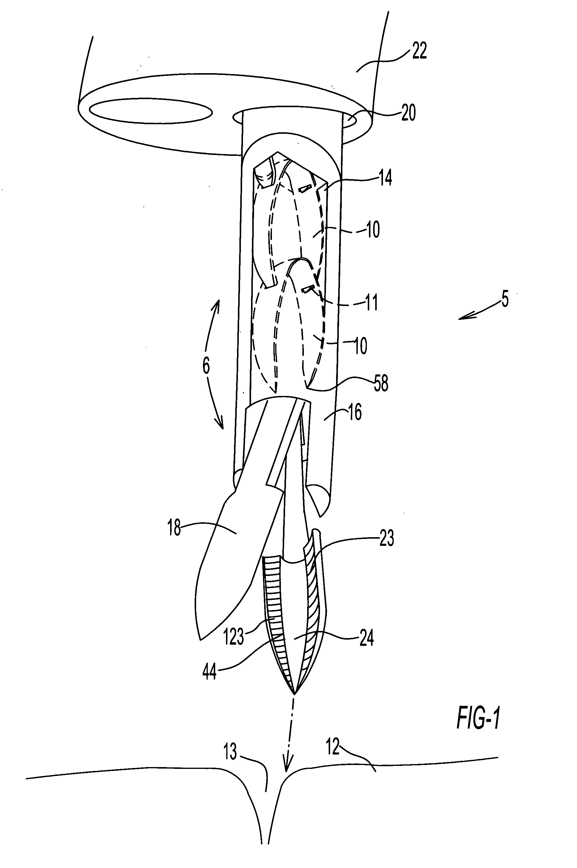
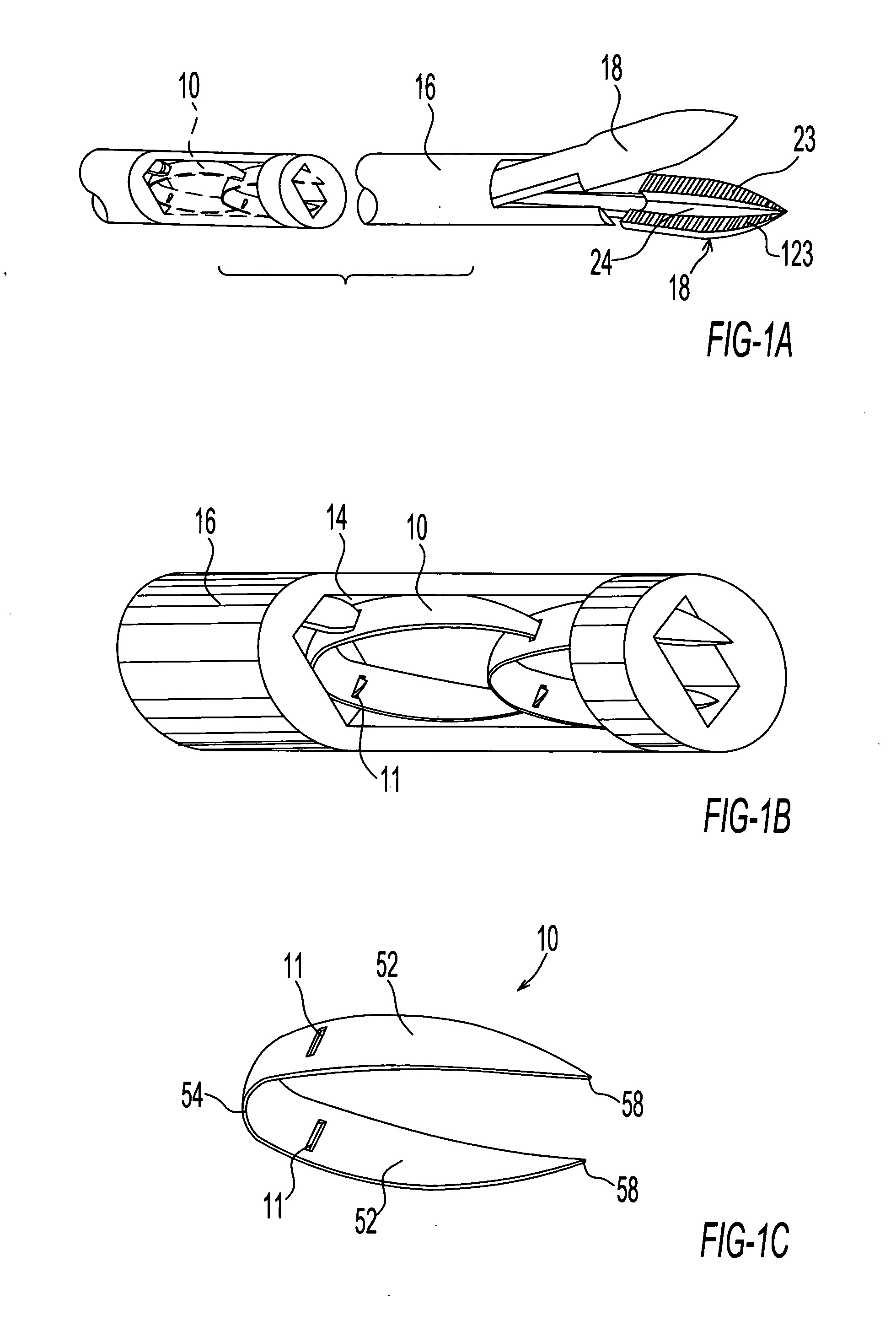
[0065] As illustrated in FIG. 1, an endoscopic fastening system 5 comprises one or more surgical fasteners 10 and a fastener delivery and deployment assembly 6, the assembly having a diameter sufficiently small so that it may be slidably insertable into an outer tubular member 20 in the form of a working channel extending longitudinally through an endoscope 22. The endoscopic fastening system, in one embodiment, for example, may be used in conjunction with an endoscope 22 having a working channel 20 2.8-4.2 mm in diameter and a shaft length of 230 cm.

[0066] Fastener delivery and deployment assembly 6 comprises a tubular shaft member 16, the shaft member being provided with a channel or lumen 14 extending longitudinally through the shaft member. Shaft member 16 is further provided at a distal end thereof with forceps jaws 18 configured for penetration of target tissue 12 and for closing fastener 10, as discussed further herein below. As shown in FIG. 1D, forceps jaws 18 are provided...


PUM


Abstract

An endoscopic fastening system having a diameter sufficiently small so that the fastening system is slidably insertable into a working channel of a flexible or rigid endoscope for performing a stapling, clipping, or other fastening operation, comprises one or more fasteners, and a fastener delivery and deployment assembly. One or more fasteners being disposed in a substantially closed prefiring configuration inside the fastener delivery and deployment system are configured for being delivered and deployed into a target tissue while remaining in a substantially closed configuration throughout the entire operation. Other related embodiments of fasteners and their respective delivery and deployment assemblies enabling fastening operations in conjunction with a flexible or rigid endoscope are described.

Description

CROSS-REFERENCE TO RELATED APPLICATIONS [0001] This application claims the benefit of the priority of U.S Provisional Application No. 60 / 557,201, filed on Mar. 29, 2004, and Provisional Application No. 60 / 611,257, filed on filed Sep. 17, 2004.FIELD OF THE INVENTION [0002] The present invention relates to an endoscopic fastening device and surgical fasteners to be used in conjunction with flexible or rigid endoscopy, or during open surgery. Fastening is performed on internal body tissues as part of a surgical diagnostic or therapeutic procedure using one or more surgical fasteners and an associated fastener delivery and deployment assembly designed for delivering one or more fasteners without having to remove the device from the body after each fastener application. BACKGROUND OF THE INVENTION [0003] Conventional surgical techniques for treating medical anomalies in the gastrointestinal tract often necessitate the use of fasteners such as, for example, staples, clips etc. Such proced...

Claims


Application Information

Patent Timeline
Patent Type & Authority Applications(United States)
IPC IPC(8): A61B17/064A61B17/068A61B17/08A61B17/10A61B17/122A61B17/128
CPCA61B17/0644A61B17/068A61B17/0682A61B17/1285A61B17/10A61B17/122A61B17/083
Inventor NAKAO, NAOMI L.
Owner GRANIT MEDICAL INNOVATION
Why Eureka
Try Eureka