
Medical treatment system, endoscope system, endoscope insert operation program, and endoscope device

a treatment system and endoscope technology, applied in the field of medical treatment system, endoscope system, endoscope insertingoperation program, endoscope apparatus, etc., can solve the problem of inability to accurately detect the gravity direction of the distal-end portion the inability to accurately reach the target portion for a short time, and the limitation of the outer diameter of the inserting portion in the endoscop

Inactive Publication Date: 2005-12-08
OLYMPUS CORP
View PDF7 Cites 94 Cited by
  • Summary
  • Abstract
  • Description
  • Claims
  • Application Information

AI Technical Summary

Benefits of technology

[0020] In addition, according to the present invention, an endoscope apparatus comprises: a detecting unit which detects inserting-operation information of an endoscope inserting portion which is inserted in a sample; a storing unit which stores standard inserting-operation information that is detected by the detecting unit and an endoscope image obtained by picking up an image of the sample taken upon the i

Problems solved by technology

In this case, in the lumen like the bronchi in the body cavity having many branches, when the abnormal portion is close to the end of the branch, the inserting portion of the endoscope does not accurately reach the target portion for a short time.
However, as disclosed in Japanese Unexamined Patent Application Publication No. 11-281897, the gravity detecting means such as the gyroscope is not provided on the distal-end portion of the inserting portion in the bronchoscope which has the limitation at the outer diameter of the inserting portion in the endoscope.
Thus, in the conventional bronchoscope apparatus, the gravity direction of the distal-end portion of the inserting portion is not accurately detected.

Method used


Image

  • Medical treatment system, endoscope system, endoscope insert operation program, and endoscope device
  • Medical treatment system, endoscope system, endoscope insert operation program, and endoscope device
  • Medical treatment system, endoscope system, endoscope insert operation program, and endoscope device

Examples


first embodiment

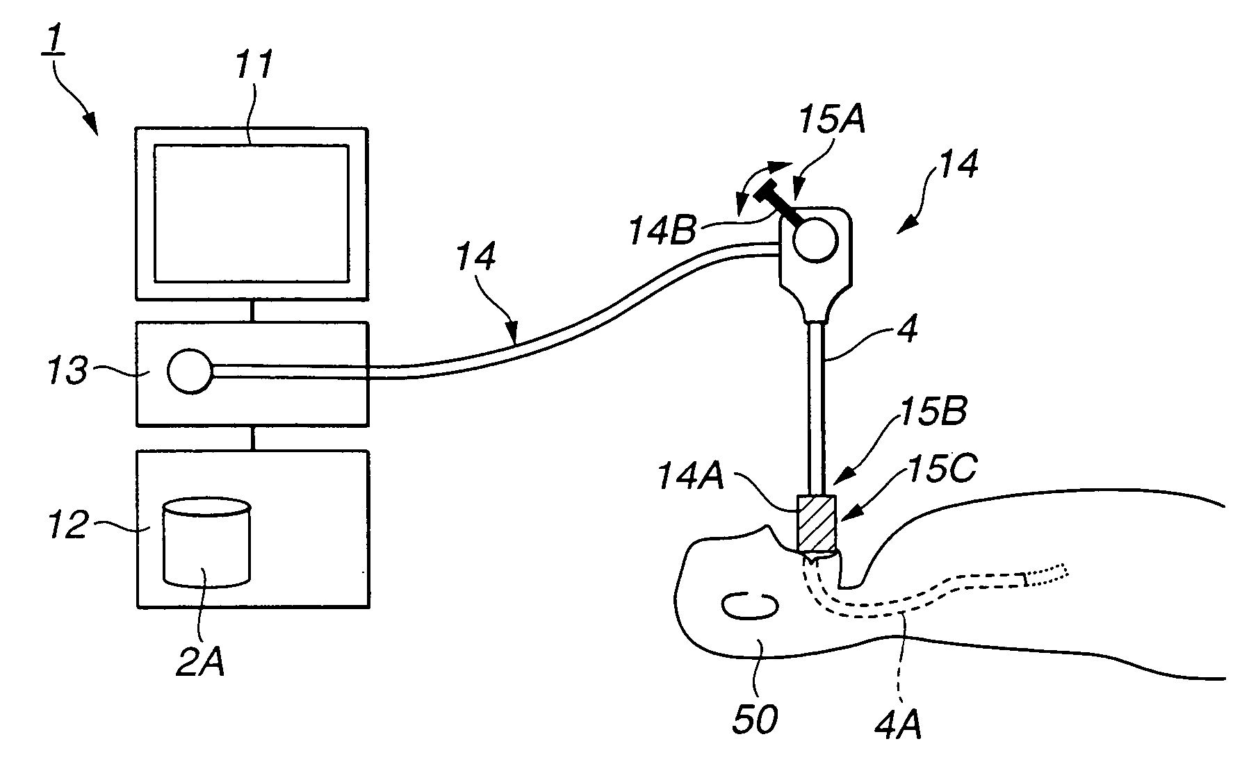
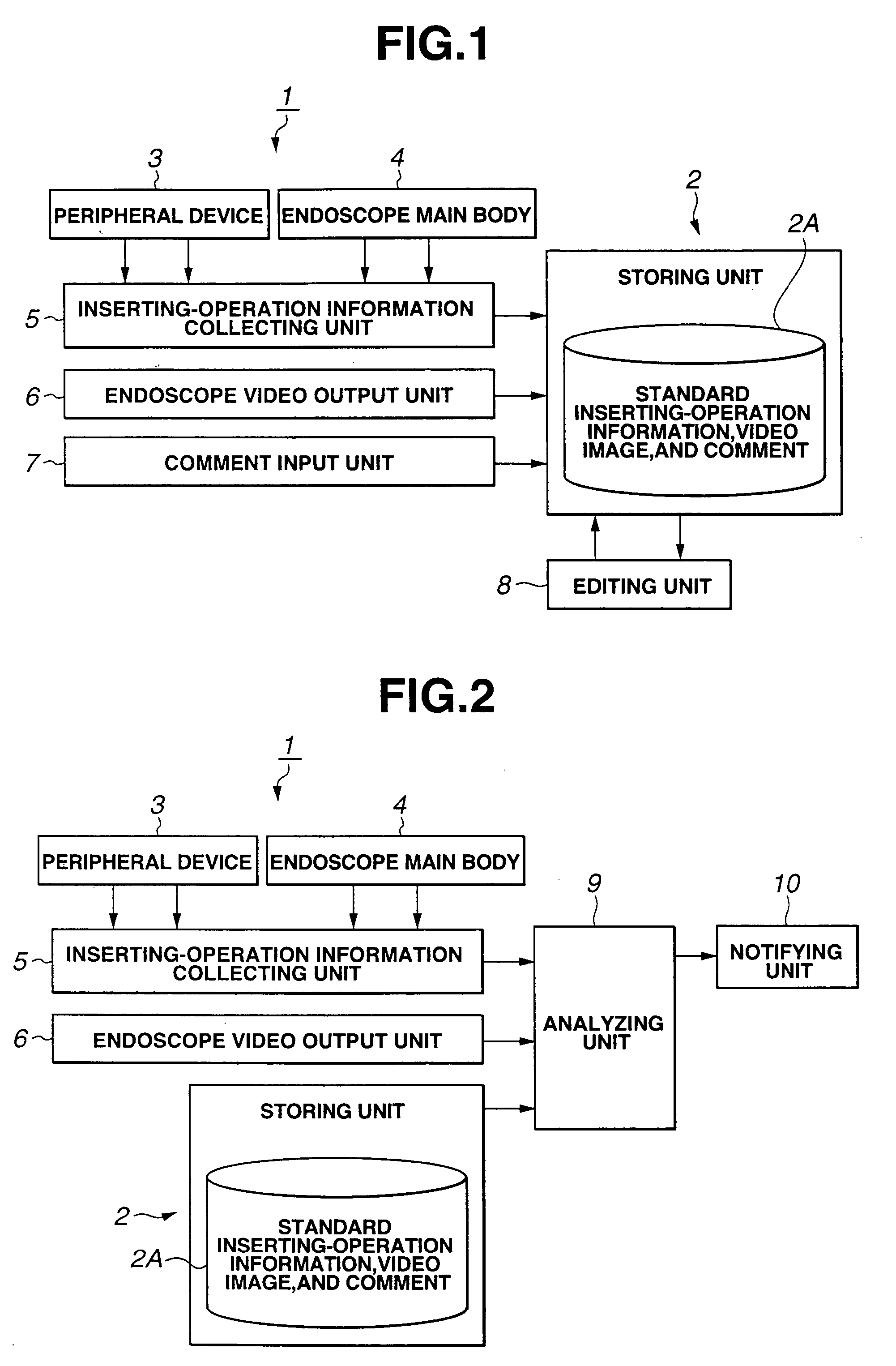
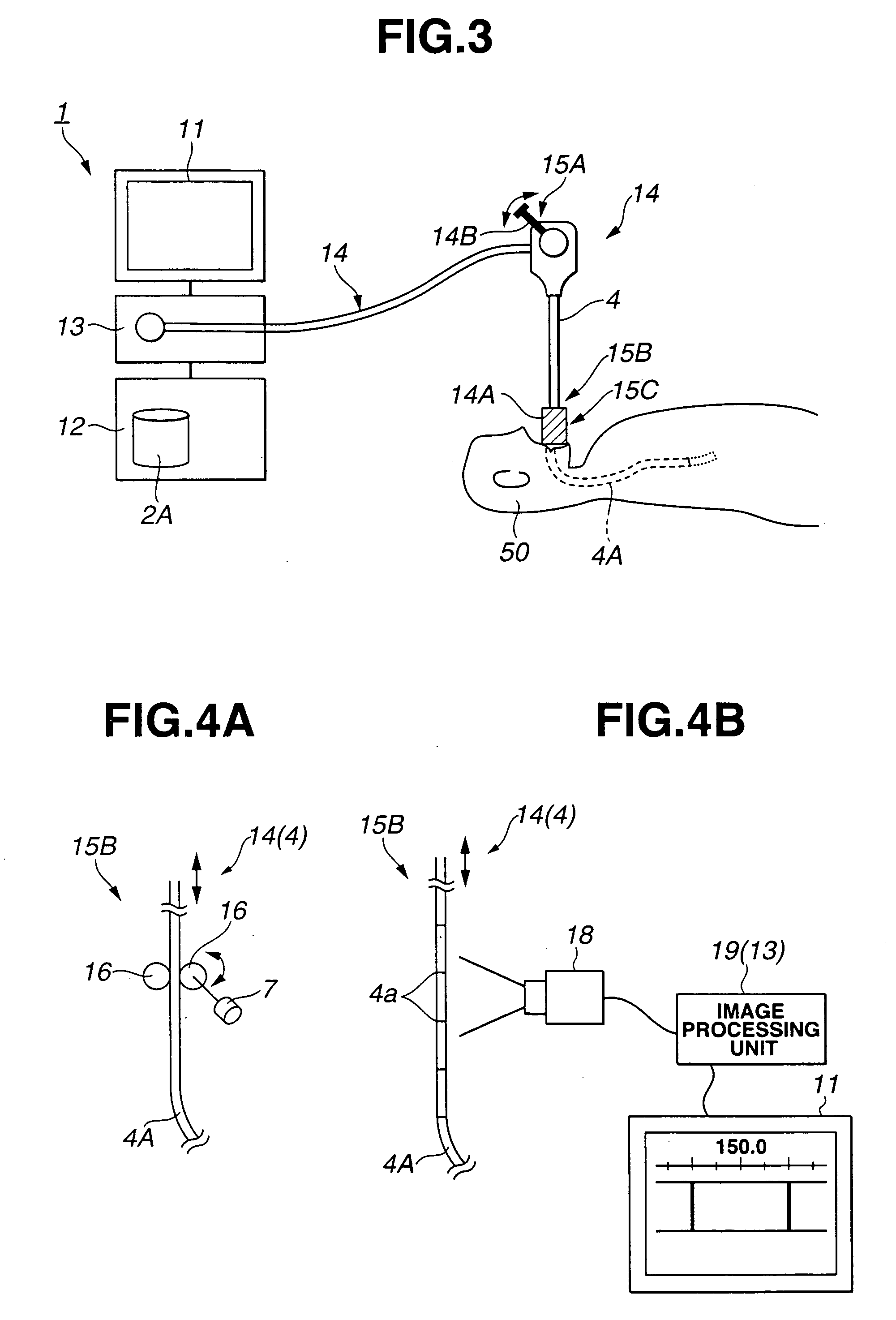
[0085] FIGS. 1 to 8 show an endoscope system and an endoscope apparatus according to the first embodiment of the present invention.

[0086] Referring to FIG. 1, an endoscope system or an endoscope apparatus 1 comprises: a processing device 2 having a storing unit 2A; a peripheral device 3 necessary for endoscope diagnosis; an endoscope main body 4 having an endoscope inserting portion as a bronchoscope, which will be described later; an inserting-operation information collecting unit 5; an endoscope video output unit 6; a comment input unit 7; and an editing unit 8.

[0087] The inserting-operation information collecting unit 5 detects and collects, as standard inserting-operation information, information of inserting action by a skilled operator who inserts the endoscope inserting portion in the bronchi. The inserting-operation information collecting unit 5 comprises a detecting unit such as a sensor which can obtain the inserting-operation information via the peripheral device 3 and ...

second embodiment

[0134] FIGS. 9 to 12 show an endoscope system or an endoscope apparatus according to the second embodiment of the present invention. Referring to FIGS. 9 to 12, the same components as those of the endoscope apparatus 1 according to the first embodiment are designated by the same reference numerals, and only different portions are described.

[0135] The endoscope system or endoscope apparatus according to the second embodiment is structured by adding an automatic inserting-operation unit 23 which automatically controls the inserting operation based on the instruction for the inserting operation using the notifying unit 10 according to the first embodiment. Other structures are the same as those of the endoscope apparatus 1 according to the first embodiment.

[0136] Referring to FIG. 9, in an endoscope apparatus (system) 1B according to the second embodiment, the automatic inserting-operation unit 23 is arranged among the analyzing unit 9, the peripheral device 3, and the bronchoscope 1...

third embodiment

[0152] FIGS. 13 to 15 show an endoscope system or the endoscope apparatus according to the third embodiment of the present invention. As shown in FIGS. 13 to 15, the same components as those according to the first embodiment are designated by the same references, a description thereof is omitted, and only different portions are described.

[0153] According to the third embodiment, in place the bronchoscope 14 according to the first embodiment, an endoscope apparatus (system) 1C comprises a training endoscope unit 31 constituting a training bronchoscope having the similar structure with the first embodiment. Further, the endoscope apparatus (system) 1C comprises a virtual endoscope image data output unit 6A which outputs virtual endoscope image data, in place of the endoscope video output unit 6, and an editing and analyzing unit 32 which can perform the similar processing of the editing unit 8 and the analyzing unit 9. Other structures are the same as those according to the first emb...


PUM


Abstract

An analyzing unit 9 is connected to an inserting-operation information collecting unit 5, an endoscope video output unit 6, and a processing device 2, and receives live inserting-operation information and endoscope image, standard inserting-operation information as stored information, and an endoscope image correlated therewith (endoscope image which is obtained by picking up an image of a sample at the position of a distal-end portion of an inserting portion detected based on the inserting-operation information). The analyzing unit 9 collects in real time the inserting-operation information and the endoscope image from the inserting-operation information collecting unit 5 and the endoscope video output unit 6 under the bronchoscope operation of an operator. Further, the analyzing unit 9 sequentially analyzes the collected inserting-operation information and endoscope image by comparing with the standard inserting-operation information and endoscope image correlated with the inserting-operation information, which are stored in a storing unit 2A of the processing device 2, outputs an analysis result to a notifying unit 10, and monitors the inserting operation of the endoscope inserting portion performed by the operator.

Description

TECHNICAL FIELD [0001] The present invention relates to a medical treatment system, an endoscope system, an endoscope inserting-operation program, and an endoscope apparatus which can perform the inserting operation of an inserting portion of, e.g., an endoscope to the lumen in the body cavity. BACKGROUND ART [0002] Recently, an endoscope system and an endoscope apparatus have widely been used. In the endoscope system and the endoscope apparatus, a long inserting portion is inserted in the body cavity to observe the organs in the body cavity, and various cure and treatments are performed by using a treatment tool which is inserted in a channel for inserting the treatment tool if needed. [0003] Further, recently, in the medical endoscope system and endoscope apparatus, a tomographic image of a sample is picked by a CT (Computed Tomography) and the diagnosis of the diseased part is performed by a three-dimensional image which is obtained from the tomographic image. [0004] One of the t...

Claims


Application Information

Patent Timeline
IPC IPC(8): A61B1/018A61B1/04A61B1/267
CPCA61B1/00009A61B1/00048A61B1/0005A61B1/00089A61B1/00101A61B1/018A61B1/042A61B1/2676A61B2090/067A61B2090/061A61B34/20A61B5/065A61B5/068
Inventor OHNISHI, JUNICHIAKIMOTO, SHUNYAKAJI, KUNIHIDEKOBAYASHI, EIICHISAITO, AKITOSHIBASAKI, TAKAOMINE, TAIJI
Owner OLYMPUS CORP
Learn More