Method and apparatus for guiding a medical instrument to a subsurface target site in a patient

Inactive Publication Date: 2006-02-16
GENERAL ELECTRIC CO
View PDF62 Cites 164 Cited by
  • Summary
  • Abstract
  • Description
  • Claims
  • Application Information

AI Technical Summary

Benefits of technology

[0007] Certain aspects of an embodiment of the present invention relate to a system and method for aiding a user in guiding a medical instrument to a target site in a patient. The system comprises an imaging device for generating one or more intraoperative images, on which spatial features of a patient target site can be defined in a 3-dimensional coordinate system. A tracking system tracks the position and optionally, the orientation of the medical instrument and imaging device in a reference coordinate system. An indicator allows a user to indicate a spatial feature of a target site on such image(s). The system also includes a display device, an electronic computer (operably connected to said tracking system, display device, and indicator), and computer-readable code. The computer-readable code, when used to control the operation of the computer, is operable to carry out the steps of (i) recording target-site spatial information indicated by the user on said image(s), (ii) determining from the spatial feature of the target site indicated on said image(s), 3-D coordinates of the target-site spatial feature in a reference coordinate system, (iii)tracking the position of the instrument in the reference coordinate system, (iv) projecting onto a display device, a view field as seen from a known position and, optionally, a known orientation, with respect to the tool, in the reference coordinate system, and (v) projecting onto the displayed view field, indicia whose states indicate the indicated spatial feature of the target site with respect to said known position and, optionally, said known orientation. Thus, the system allows the user, by observing the states of said indicia, to guide the instrument toward the target site by moving the instrument so that said indicia are placed or held in a given state in the displayed field of view.

Problems solved by technology

These procedures generally require the surgeon to operate on portions of the anatomy that are not directly visible, or can be seen only with difficulty.
Furthermore, some parts of the body contain extremely complex or small structures and it is necessary to enhance the visibility of these structures to enable the surgeon to perform more delicate procedures.

Method used


Image

  • Method and apparatus for guiding a medical instrument to a subsurface target site in a patient
  • Method and apparatus for guiding a medical instrument to a subsurface target site in a patient
  • Method and apparatus for guiding a medical instrument to a subsurface target site in a patient

Examples


Embodiment Construction

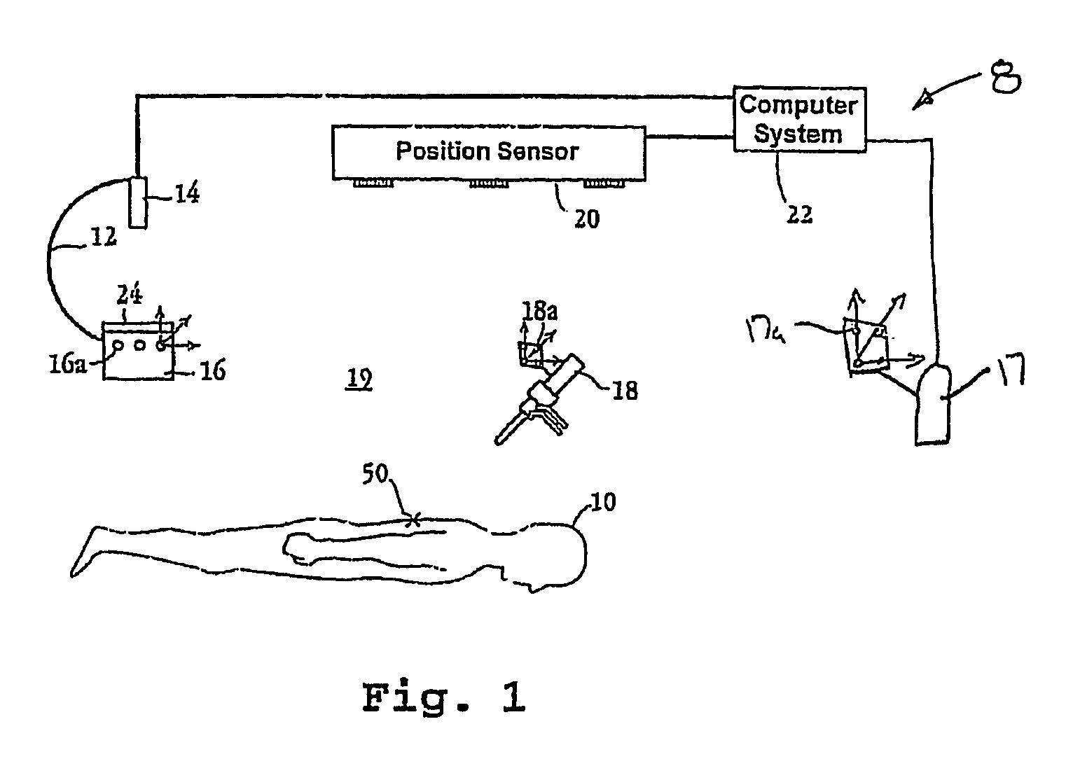
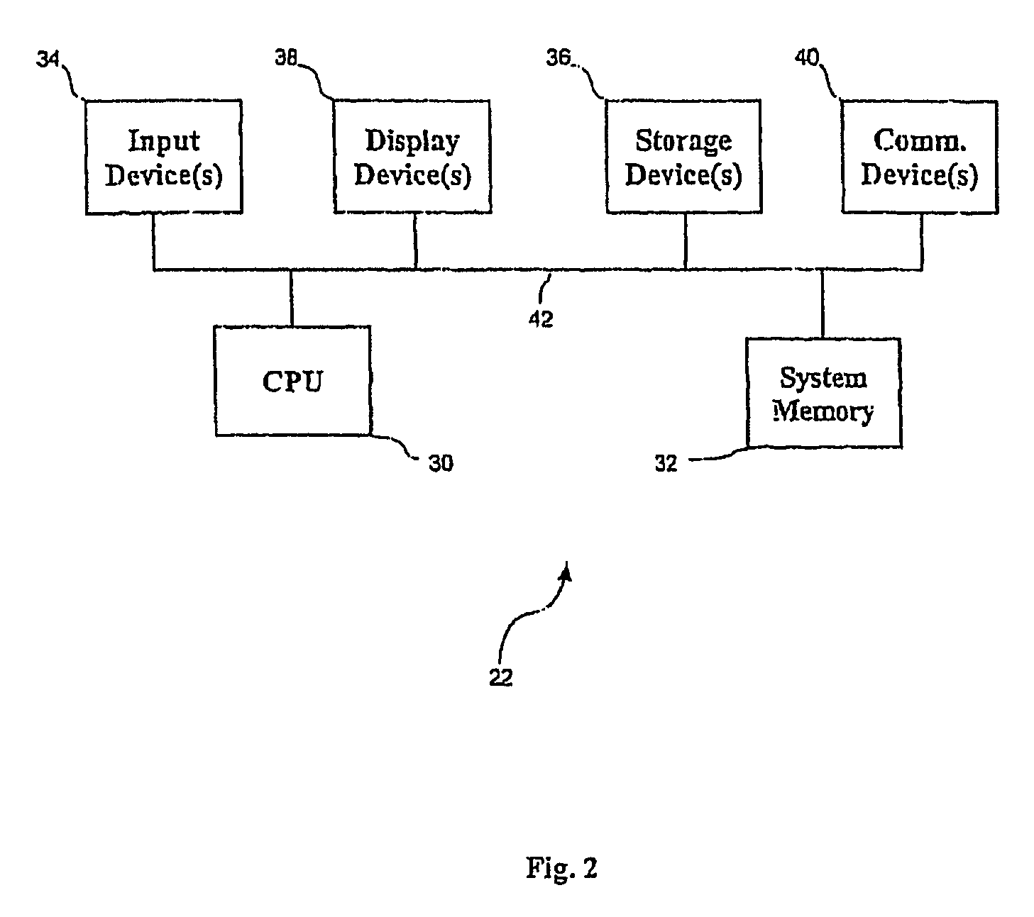
[0035]FIG. 1 is a schematic view of an image-guided surgery system 8 according to certain aspects of an embodiment of the invention. The system includes an imaging device for generating intraoperative images of selection portions of the patient's 10 anatomy. For example, as shown in FIG. 1 the imaging device may comprise a mobile fluoroscopic device 12. Fluoroscopic device 12 is preferably a C-Arm of the type which may be obtained from General Electric, Milwaukee, Wis. The mobile fluoroscopic device includes an X-ray camera 14 and an image intensifier 16. Alternatively, the imaging device may be an ultrasound imaging device, such as a hand held ultrasound imaging probe 17. The system also includes a surgical instrument 18, which may be any of a variety of devices such as a pointer, a drill, or an endoscope, for example. The system also includes a tracking system. In this respect, the C-arm / image intensifier 24, the ultrasound probe 17 and the surgical instrument 18 are each equipped...


PUM


Abstract

Intraoperative image(s) of a patient target site are generated by an intraoperative imaging system (e.g., ultrasound or X-ray). The intraoperative imaging system is tracked with respect to the patient target site and surgical instrument(s) (e.g., a pointer, endoscope or other intraoperative video or optical device). The intraoperative images, surgical instruments, and patient target site are registered into a common coordinate system. Spatial feature(s) of the patient target site are indicated on the images of the patient target site. Indicia relating the position and orientation of the surgical instrument(s) to the spatial feature(s) of the patient target site are projected on the images, with the indicia being used to correlate the position and orientation of the surgical instruments with respect to the target feature.

Description

RELATED APPLICATIONS [0001] This application makes reference to and claims priority from U.S. Provisional Patent Application Ser. No. 60 / 541,131 entitled “Method and Apparatus for Guiding a Medical Instrument to a Subsurface Target Site in a Patient” filed on Feb. 2, 2004, the complete subject matter of which is incorporated herein by reference in its entirety.FEDERALLY SPONSORED RESEARCH OR DEVELOPMENT [0002] [Not Applicable]MICROFICHE / COPYRIGHT REFERENCE [0003] [Not Applicable]BACKGROUND OF THE INVENTION [0004] Precise imaging of portions of the anatomy is an increasingly important technique in the medical and surgical fields. In order to lessen the trauma to a patient caused by invasive surgery, techniques have been developed for performing surgical procedures within the body through small incisions with minimal invasion. These procedures generally require the surgeon to operate on portions of the anatomy that are not directly visible, or can be seen only with difficulty. Further...

Claims


Application Information

Patent Timeline
Patent Type & Authority Applications(United States)
IPC IPC(8): A61B5/05
CPCA61B5/06A61B19/5212A61B19/5244A61B2019/5289A61B2019/5238A61B2019/5255A61B2019/5276A61B2019/507A61B5/062A61B5/064A61B34/20A61B90/361A61B2034/107A61B2034/2055A61B2090/364A61B2090/376A61B2090/378
Inventor SHAHIDI, RAMINMAURER, CALVIN R.WEST, JAYKHADEM, RASOOL
Owner GENERAL ELECTRIC CO
Why Eureka
Try Eureka