System and method for marking an anatomical structure in three-dimensional coordinate system

a three-dimensional coordinate system and anatomical structure technology, applied in the field of ultrasound tracking systems, can solve the problems of difficult to navigate the catheter to the appropriate location, difficult for the physician to be certain of the catheter position, and inability to endoscopic visualization of the treatment site within the body, etc., to facilitate the navigation of the medical device(s) within the patient.

Inactive Publication Date: 2006-03-16
BOSTON SCI SCIMED INC
View PDF70 Cites 50 Cited by
  • Summary
  • Abstract
  • Description
  • Claims
  • Application Information

AI Technical Summary

Benefits of technology

[0009] The present invention is a device localization system that uses one or more ultrasound reference catheters to establish a fixed three-dimensional coordinate system within a patient's heart, preferably using principles of triangulation. The coordinate system is represented graphically in three-dimensions on a video monitor and aids the clinician in guiding other medical devices, which also carry ultrasound transducers, through the body to locations at which they are needed to perform clinical procedures. In one embodiment of a system according to the present invention, the system is used in the heart to help the physician guide mapping catheters for measuring electrical activity, and ablation catheters for ablating selected regions of cardiac tissue, to desired locations within the heart.
[0010] Three-dimensional images are shown on a video display which represent the three-dimensional positions and orientations of at least portions of the medical devices used with the system, such as the reference catheter, and the electrodes of the mapping catheter and ablation catheter. The video display may additionally include representations of the electrical activity measured by each mapping electrode at its respective location on the three-dimensional display. It may also represent ablation lesions formed within the body at the appropriate three-dimensional locations, and / or certain anatomic structures which may facilitate navigation of the medical device(s) within the patient.

Problems solved by technology

For certain types of minimally invasive medical procedures, endoscopic visualization of the treatment site within the body is unavailable or does not assist the clinician in guiding the needed medical devices to the treatment site.
Because the fluoroscopic image is in two-dimensions and has fairly poor resolution, it may be difficult for the physician to be certain of the catheter positions.
Thus, for example, once a physician has identified an area which is to be ablated (using a map of the measured electrical activity of the heart) it may be difficult to navigate an ablation catheter to the appropriate location in order to accurately ablate the area of concern.
While useful, neither of the disclosed systems provides for determining medical device locations relative to a fixed coordinate system within the body.
The lack of a fixed coordinate system within the body can lead to tracking errors which in turn render it difficult to guide medical devices to the desired locations within the body.

Method used


Image

  • System and method for marking an anatomical structure in three-dimensional coordinate system
  • System and method for marking an anatomical structure in three-dimensional coordinate system
  • System and method for marking an anatomical structure in three-dimensional coordinate system

Examples


Embodiment Construction

Localization System Overview

[0052] The localization system and procedure will next be described in general terms. Specific examples of procedures which may be carried out using the system will be described in the Operation section of this description. The system is described primarily with respect to catheters in the heart, but it should be understood that the system is intended for use with other medical devices and in other regions of the body as well.

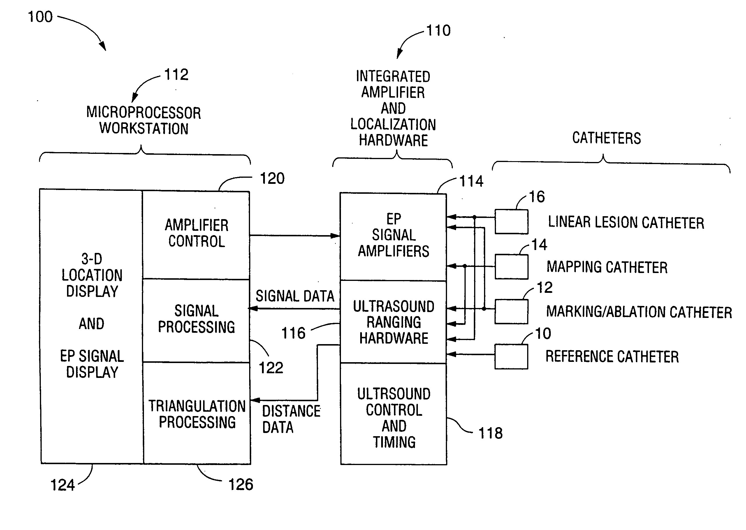
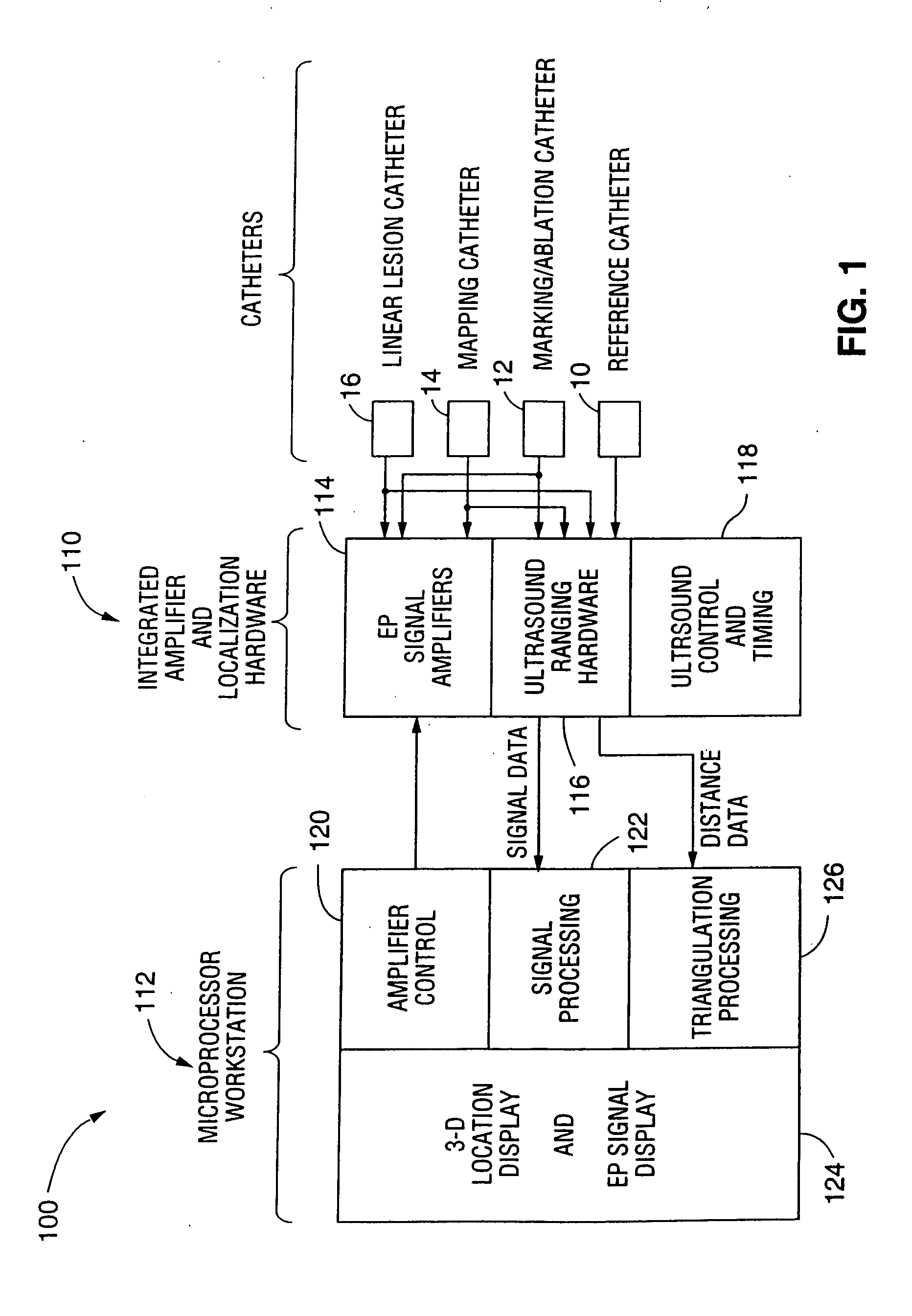
[0053] Referring to FIG. 1, the present invention is a device localization system 100 that uses one or more ultrasound reference catheters 10 to establish a three-dimensional coordinate system within a patient's heart. The system allows the positions of one or more additional catheters 12, 14, 16, to be represented graphically on a graphical user interface 124 relative to a coordinate system. This aids the clinician in guiding the additional catheter(s) 12, 14, 16 through the heart to locations at which they are needed to perform ...


PUM

PropertyMeasurementUnit
velocityaaaaaaaaaa
diameteraaaaaaaaaa
diameteraaaaaaaaaa

Abstract

The present invention is a device localization system that uses one or more ultrasound reference catheters to establish a fixed three-dimensional coordinate system within a patient's heart using principles of triangulation. The coordinate system is represented graphically in three-dimensions on a video monitor and aids the clinician in guiding other medical devices, which are provided with ultrasound transducers, through the body to locations at which they are needed to perform clinical procedures. In one embodiment of a system according to the present invention, the system is used in the heart to help the physician guide mapping catheters for measuring electrical activity, and ablation catheters for ablating selected regions of cardiac tissue, to desired locations within the heart.

Description

FIELD OF THE INVENTION [0001] The present invention relates generally to the field of ultrasound tracking systems. More specifically, it relates to systems for tracking the positions of devices within the human body. BACKGROUND OF THE INVENTION [0002] For certain types of minimally invasive medical procedures, endoscopic visualization of the treatment site within the body is unavailable or does not assist the clinician in guiding the needed medical devices to the treatment site. [0003] Examples of such procedures are those used to diagnose and treat supra-ventricular tachycardia (SVT), atrial fibrillation (AF), atrial flutter (AFL) and ventricular tachycardia (VT). SVT, AFL, AF and VT are conditions in the heart which cause abnormal electrical signals to be generated in the endocardial tissue to cause irregular beating of the heart. [0004] A procedure for diagnosing and treating SVT or VT involves measuring the electrical activity of the heart using an electrophysiology catheter int...

Claims


Application Information

Patent Timeline
Patent Type & Authority Applications(United States)
IPC IPC(8): A61B8/00A61B5/042A61B5/06A61B8/08A61B8/12A61B18/14
CPCA61B5/0422A61B8/445A61B5/6855A61B5/6856A61B5/6858A61B8/0833A61B8/12A61B8/543A61B18/1492A61B2017/00106A61B2018/00065A61B2018/00113A61B2018/00214A61B2018/00351A61B2018/00577A61B2018/00839A61B2019/5251A61B2019/5263A61B2218/002A61B2562/0209A61B2562/043A61N2007/0078A61B5/06A61B2034/2051A61B2034/2063A61B2562/0215A61B5/287
Inventor WILLIS, N. PARKERBRISKEN, AXELZENG, JINGLINHURD, MARSHA
Owner BOSTON SCI SCIMED INC
Why Eureka
Try Eureka